need help with your account or subscription? click here to email us (or see the contact page)
join telegramNEW! discord
jump to exam page:
search for anything โ‹… score predictor (โ€œpredict me!โ€)

Welcome to peridotโ€™s page.
Contributor score: 115


Comments ...

 +6  visit this page (nbme24#20)
get full access to all content โ‹… become a member

Paraxial mesoderm means mesoderm surrounding the axis, aka the neural tube. They eventually form a pair of somites on either side. Somites then differentiate into:

  • sclerotomes -> vertebrae, rib cage, occipital bone (part)
  • myotomes -> skeletal muscle of the back, ribs, limbs
  • syndetomes -> cartilage, tendons
  • dermatomes -> skin of the back

Source

In this case, the patient is lacking spinous processes on top of having spina bifida. Maybe you were thinking spina bifida = neuropore defect, but what does this have to do with sclerotomes? This is what I found:

"Failure of closure of the caudal neuropore during embryogenesis will lead to spina bifida. This condition is always marked by a local lack of osteogenesis, or bone growth. The reason is that the correct differentiation and placement of underlying tissues (the spinal chord and spinal nerves and associated tissues) induce the osteogenesis, even small flaws in the underlying tissues can lead to a problem in the formation of the spine (vertebrae)." Source

So I'm guessing that when the question asks for "cause of the defect" they are talking about the vertebrae, not exactly the cause of spina bifida, which would be failure of neuropores to fuse. If that was an answer choice then def pick that, but the lack of vertebrae is due to lack of sclerotome fusion (because spina bifida could not induce it properly).

get full access to all content โ‹… become a member

 +14  visit this page (nbme24#23)
get full access to all content โ‹… become a member

In case anyone else was wondering about the other choices:

A: Acetyl-CoA carboxylase is the first committed step in fatty acid synthesis

B: Homocysteine methyltransferase turns homocysteine into methionine (we can deduce this from knowledge about homocystinuria)

C: Methionine adenosyltransferase makes SAM. SAM has two roles that we know of - one in turning methionine back into homocysteine, and one turning norepinephrine into epinephrine. This could make it a tempting choice but at the time I just saw the word 'methionine' and thought that couldn't have anything to do with cortisol/catecholamines.

D: LOL for methylmalonyl-CoA racemase, wiki says "It is routinely and incorrectly labeled as 'methylmalonyl-CoA racemase'. It is not a racemase because the CoA moiety has 5 other stereocenters." Good job NBME. Anyway, methylmalonyl-CoA epimerase is involved in the same catabolic pathway that we know of through propionic acidemia, so it's involved in breakdown of fatty acids and amino acids.

get full access to all content โ‹… become a member
jurrutia  SAM is also the methyl donor used by DNA methyltransferase (DNa methylation) +

 +12  visit this page (nbme21#15)
get full access to all content โ‹… become a member

Here my stab at summarizing all the info. The first diagram on this website is a super helpful as well; This diagram sorta helps too.

Normally, bilirubin from the blood stream is conjugated in the liver to a water-soluble form. This gets mixed with bile to be excreted through the intestines. In the small intestine, it becomes urobilinogen, some of which is excreted by the kidneys, giving pee that good yellow color. Some of the urobilinogen turns into stercobilin and stays in the GI to be excreted in poop, giving poop that good brown color.

In bile duct obstruction, bilirubin gets conjugated to a water-soluble form - then it's stuck. It's unable to be excreted into the intestines, unable to turn into urobilinogen or stercobilin, etc. Conjugated bilirubin builds up in the liver, and eventually gets backed up back into the blood stream where it came from. Since it's water-soluble and floating around in circulation, it eventually gets filtered by the kidneys and ends up in pee. Bilirubin is darker in color, so the pee would be dark. However, the stool would be pale since no stercobilin was made. I believe that symptoms of itchy skin also occurs in these cases as bile salts back up and are deposited in skin.

Hemolysis is not the correct answer because most of the bilirubin is not yet conjugated, so it's not water-soluble and doesn't end up in the kidneys.

get full access to all content โ‹… become a member
notpennysboat  Amazing explanation, thanks a lot! +2

 +1  visit this page (nbme21#41)
get full access to all content โ‹… become a member

SPOILER ALERT!!

Do not read ahead if you have not taken NBME 22!!!


I'm smh at this question because in NBME 22, they describe a scenario in which loperamide caused CNS effects - particularly ventilatory depression and we were supposed to know that it was loperamide. I guess this question specified "little to no" rather than "none at all" but come ON, I'm annoyed that we have to separate such nitpicky detail when one question emphasizes its lack of CNS effects, then they turn around and test us on an exception of this exact same content. /end rant

This was not very informative at all, I just wanted to consolidate a similar question that I knew I had just seen but it turned into me just being angry I'm sorry.

get full access to all content โ‹… become a member

 +6  visit this page (nbme21#8)
get full access to all content โ‹… become a member

I am for some reason learning about this for the first time so I'll write a bit about euthyroid sick syndrome in case it helps someone.

It's a disease of exclusion - the thyroid hormones are whack but the thyroid gland itself seems to be totally fine. Specifically, T3 levels are low, rT3 is high (the activity of different deiodinases are off, so rT3 is made more and degraded less, though it doesn't seem like the rT3 level has any other clinical implications). As the other comments have mentioned, TSH and T4 are typically normal, although they can be decreased as well in severe cases.

Euthyroid sick syndrome is typically due to an underlying critical illness or starvation - the body stops making the hormones as an extreme measure to save energy and resources. The treatment is to treat the underlying illness; thyroid hormone therapy is not recommended as its effectiveness is inconclusive.

In this question stem, the patient has an underlying illness. Her thyroid gland works fine (responds appropriately to TSH). Classic case of euthyroid sick syndrome.

Given that her T4 levels are normal, we can rule out B, D, and E. I'm a dumdum and put C because I had no idea what euthyroid sick syndrome was. But given that her TSH level is also normal, it's not an issue with the hypothalamus or pituitary gland.

(Sources: wiki page and this article)

get full access to all content โ‹… become a member
schep  Thanks! I, too, am hearing about this for the first time +

 +11  visit this page (nbme21#34)
get full access to all content โ‹… become a member

Here is my summary of the picture/video that was posted:

There are 3 pathways involved in peeing:

  1. Pelvic n. (aka pelvic splanchnic n.) sends parasympathetic fibers to deltrusor to contract --> squeeze bladder and pee.

  2. Hypogastric n. sends sympathetic fibers to the deltrusor to relax, as well as the internal sphincter to contract --> hold back pee

  3. Pudendal n. sends somatic fibers (under conscious control) to the external sphincter to contract --> hold back pee

In this question, the patient's bladder is filling up so much that it's forced to overflow. That means there is a problem with scenario 1 - damage to pelvic n. so that he can't squeeze his bladder even when it's super full.

get full access to all content โ‹… become a member
peridot  To clarify, this description is meant to go along with @hungrybox's pic link and @eacv's video link +4
lovebug  thanks a lot! +

 +0  visit this page (nbme22#22)
get full access to all content โ‹… become a member

While I understand why it's hyperplastic arteriosclerosis and how it classically occurs with HTN, I was wondering why it couldn't be berry aneurysm? Is it because the question is asking which is "most likely", making C the better answer? Thank you.

get full access to all content โ‹… become a member
mannan  Berry Aneurysm is not CAUSED by HTN. It's caused by weakening of the arterial wall (at bifurcations). Hypertensive disease exacerbates them and causes the clinical picture of SAH (worst headache of life) when they rupture. Hope that helps -- Reference: FA CNS pathology, aneurysms. +2
usmleboy  Actually according to Goljan this is incorrect. Berry aneurysms are caused by hypertension. The weakening of the wall (no tunica media) at bifurcations is inherent in human anatomy. Basically you have to have elevated BP to cause the dilation, outside of the inherited connective tissue disorders. Hence why PKD has the berry association. However, these aneurysms present with extremely prolonged HTN, whereas our guy in this Q only has a 1 year history. The key to answering this question is recognizing that this is MALIGNANT hypertension that is relatively acute in onset. Malignant HTN = hyperplastic arteriolosclerosis (onion rings). +3


Subcomments ...

submitted by b1ackcoffee(115), visit this page
get full access to all content โ‹… become a member

from @melchior

From the UW ID 666 explanation, although type II pneumocytes normally differentiate into type I pneumocytes after proliferation, they do not differentiate in idiopathic pulmonary fibrosis due to altered cell signals and altered basement membrane, which is why type II pneumocytes are increased.

explanation by @benwhite_dotcom is incorrect

get full access to all content โ‹… become a member
peridot  The UWorld example is talking about idiopathic pulmonary fibrosis, whereas benwhite_dotcom was talking about chronic inflammatory pneumonitis, which presents with increased type II pneumocytes (as indicated by the correct answer). +3
topgunber  either way destruction of basement membrane leads to type II hyperplasia. It may leads to fibrosis because of destruction of the basement membrane (the point of no return for lung parenchyma). Type I pneumoyctes will not rise in number because the destruction in the basement membrane (they are the vast majority of cells covering alveoli) +3


submitted by misterdoctor69(70), visit this page
get full access to all content โ‹… become a member

Damn everyone out here looking at the eyes when my dumbass was thinking the girl was missing a nasal bridge or something lol fml

get full access to all content โ‹… become a member
peridot  That is straight up what I wrote: "low nasal bridge???" I was like is this part of some congenital defect +
mkayman  lmfao +


submitted by colonelred_(124), visit this page
get full access to all content โ‹… become a member

The analysis only showed a mutation in one allele. CF is an autosomal recessive disease: the disease only manifests if there are mutations in both alleles of the CFTR gene.

If you still have 1 functional copy of the CFTR gene, you can still make the CFTR protein (the chloride channel/transporter), hence your body wonโ€™t have any issues.

This is analogous to tumor suppressor genes like Rb: so long as one of the alleles you have is functional, you can make enough of the protein to โ€œmake upโ€ for the defective allele. If both get knocked out (Rb-/-), you lose the protection provided by the gene because now you make no protein at all.

The only thing that made sense for this question was the fact that the other allele was not included in the analysis.

get full access to all content โ‹… become a member
charcot_bouchard  OR another allele has a diff type of mutation because CF is done by like hundreds of diff type of mutation. SO the 70 types that we screened covered one type from one parent but not another that was inherited from other parent. +46
soph  I put D thinking there was a mutation in another protein that interacts with CFTR....thus u dont have CF but some disease with similar phenotype. Is this wrong bc its simply not the case ?? +14
nbmehelp  @charcot_bouchard I think that makes more sense if I understand what you're saying- Probably had a mutation only in 1 of 2 of the same alleles in the analysis but had another mutation in 2 of 2 alleles at a different location not included in the analysis, right? +
fallot4logy  CF is a rare disease , and the possibility to have a mutated gene plus a gene that its not belong to 70 most common cf mutations is extremely rare +5
gubernaculum  @soph i picked D too but now looking back, the panel had 70 of the most common CFTR gene mutations so it is unlikely that they didn't already check a gene that codes for a protein that interacts with CFTR? that's the only way i can rationalize it. its bad writing ultimately +
peridot  I also picked D, but there are over 1700 different mutations for CF and it's too hard to test for them all - the panel in the question tested for the 70 most common. As others mentioned, CF is an autosomal recessive disease, so there must be another mutated allele here for the child to present with the disease. It's more likely, and I imagine not uncommon, that the mutation is not in the panel. As for D, I suppose the best reasoning I can come up for it is that nothing like that exists - what protein interacts with ONE mutated CFTR allele in that it results in the same phenotype as CF, a disorder that requires TWO mutated alleles? I have never heard of such a thing, whereas I have definitely heard of A being the case. +2
weirdmed51  @peridot , โ€˜Bโ€™ being the cause you mean +


submitted by lsmarshall(465), visit this page
get full access to all content โ‹… become a member

MHC class 1 peptide antigen processing > "Antigen peptides loaded onto MHC I in RER after delivery via TAP (transporter associated with antigen processing)" - First Aid 2019.

Bare lymphocyte syndrome type 2 (BLS II; affecting MHC II) is due to mutations in genes that code for transcription factors that normally regulate the expression (gene transcription) of the MHC II genes. Bare lymphocyte syndrome type 1 (BLS I; affecting MHC I), is much more rare, and is associated with TAP deficiencies.

get full access to all content โ‹… become a member
tyrionwill  in the question, it says absence of MHC-I presenting cells. I guess the meaning is lack of MHC-I. IF TAP is missing or dysfunction (bare lym syn type-1), MHC-I should be there, however Ag cannot be loaded to the MHC-I. Can anyone help me to understand more. +1
peridot  @tyrionwill From wiki: "The TAP proteins are involved in pumping degraded cytosolic peptides across the endoplasmic reticulum membrane so they can bind HLA class I. Once the peptide:HLA class I complex forms, it is transported to the membrane of the cell. However, a defect in the TAP proteins prevents pumping of peptides into the endoplasmic reticulum so no peptide:HLA class I complexes form, and therefore, no HLA class I is expressed on the membrane. Just like BLS II, the defect isn't in the MHC protein, but rather another accessory protein." +1
j44n  i hate this question because MHCI is on all nucleated cells. So this person is literally a bag of RBC's +1
soccerfan23  @j44n Not quite. It's true that MHC I is on all nucleated cells. Because of the TAP mutation, these cells don't express MHC I on their membranes. But these cells still exist. That is what is meant when the vignette says "flow cytometry shows absence of class I MHC-expressing cells. +1


submitted by andro(269), visit this page
get full access to all content โ‹… become a member

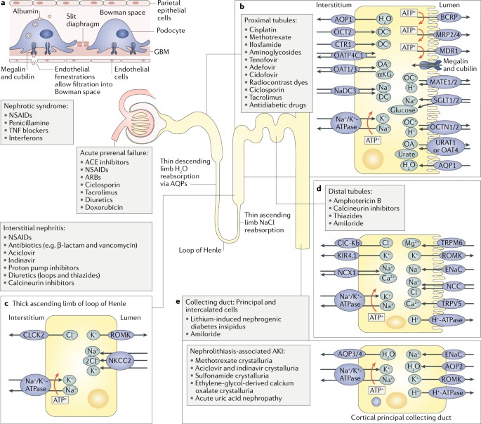
ATN ( Acute Tubular necrosis ) may be either ischemic or nephrotoxic:

  • Ischemic - tends to affect the Proximal straight segment PST and the TAL ( thick ascending limb)

*Toxic - tends to affect the PCT ( convoluted segment )

get full access to all content โ‹… become a member
peridot  this is on p.591 of FA 2019 for anyone interested +1


submitted by neonem(630), visit this page
get full access to all content โ‹… become a member

Aminoglycosides are nephrotoxic; nephrotoxic chemicals/drugs cause acute tubular necrosis (ATN), characterized by damage to the PCT. ATN causes the formation of brown, muddy, granular casts in the urine. The fact that this patient is a quadriplegic might be suggesting that they have a lower volume of distribution for the drug (and therefore higher blood concentrations).

get full access to all content โ‹… become a member
mtkilimanjaro  I would also like to add ATN is nephrotoxic ischemia and the two places in the tubule that are susceptible are the PCT (proximal straight part) and the thick ascending limb. The TAL is not labeled as a choice so that is why it has to be B (and why B is a little further down from the convoluted part) +4
mtkilimanjaro  Actually aminoglycosides might only affect the PCT idk :( +2
peridot  on p. 591 of FA 2019, it talks about ATN. The two types are 1. ischemic - affects PCT and thick ascending limb because those two areas use ATP the most (think of all the ion pumps) and 2. nephrotoxic - PCT only (I think of it as that's the first part, so it's most exposed to toxins). Aminoglycosides fall under scenario 2. +3
cassdawg  If you wanna see nephrotoxic drugs in one place, here's an image with the locations of different nephrotoxic drugs: https://media.springernature.com/lw685/springer-static/image/art%3A10.1038%2Fs41581-018-0003-9/MediaObjects/41581_2018_3_Fig1_HTML.jpg +3
corndog  Before anyone looks at @cassdawg link, consider taking some Loperamide. +6
weirdmed51  @cassdawg do you want us to be admitted in asylum before sitting for the exam? +1


submitted by andro(269), visit this page
get full access to all content โ‹… become a member

SIADH is characterized by :

*Excessive free water retention ( less water in urine means specific gravity increases and not decreases as in option E)

*Euvolemic hyponatremia with continued urinary Na+ excretion ( and so urine Na which actually be greater than and not less than 10 mEq/L option D)

*Urine osmolality > serum osmolality ( option C)

Serum potassium does not actually decrease ( counter-intuitively ) because the excess fluid retention suppresses aldosterone secretion . The two main stimuli for potassium loss/secretion in kidneys are - aldosterone - and high urine flow rates. Both of these are decreased in SIADH ( excluding option B )

Serum urea nitrogen and creatinine are diluted , so their concentration decreases

get full access to all content โ‹… become a member
peridot  Great explanation, thank you so much!! +1


submitted by m-ice(370), visit this page
get full access to all content โ‹… become a member

In SIADH, the excessive ADH causes the collecting duct of the kidney to reabsorb huge amounts of water that it should normally excrete. That means that the plasma will now have much more water relative to solute (low osmolality) and the urine will have much more salt relative to water (higher osmolality).

get full access to all content โ‹… become a member
frijoles  So potassium does not become diluted in SIADH? +1
ruready4this  I feel like I was overthinking this question so much for some reason!! C definitely makes the most sense but I was also wondering what would happen to potassium. Then I was thinking maybe the excess ADH would suppress aldosterone secretion and serum potassium concentration would actually be higher +1
peridot  @frijoles Aldosterone can adjust the K+ levels: too much water --> less aldosterone --> no excretion of K+, so this helps retain the K+ to a normal level. However, less aldosterone also means --> more excretion of Na+, so the hyponatremia is not corrected. +1


get full access to all content โ‹… become a member

Baby developed RDS --> guven O2 as Rx --> complications of O2 herapy in neonated = RIB (FA 2019) R = Retinopathy of prematurity I = Inraventricular hemorrhage B = Bronchopulmonary dysplasia

get full access to all content โ‹… become a member
peridot  RDS (respiratory distress syndrome) and O2 therapy complications are described on p.647 in FA 2019! +2
peridot  This is due to free radicles, which is also briefly touched upon on p. 210 of FA 2019 +
peridot  radicals* +1
j44n  also I saw on one of the other NBME's that retinopathy is more likely than the intraventricular hemorrhage. I did some reading and found that the eyes adapt to local changes faster. So when you give them 100%02 their eyes get used to the elevation in 02 so when you take them off of it they upregulate a ton of VEGF to try to normalize back to the level they were used to on the 100% +3
jbrito718  Stevie Wonder is a victim of retinopathy of prematurity +3


get full access to all content โ‹… become a member

Baby developed RDS --> guven O2 as Rx --> complications of O2 herapy in neonated = RIB (FA 2019) R = Retinopathy of prematurity I = Inraventricular hemorrhage B = Bronchopulmonary dysplasia

get full access to all content โ‹… become a member
peridot  RDS (respiratory distress syndrome) and O2 therapy complications are described on p.647 in FA 2019! +2
peridot  This is due to free radicles, which is also briefly touched upon on p. 210 of FA 2019 +
peridot  radicals* +1
j44n  also I saw on one of the other NBME's that retinopathy is more likely than the intraventricular hemorrhage. I did some reading and found that the eyes adapt to local changes faster. So when you give them 100%02 their eyes get used to the elevation in 02 so when you take them off of it they upregulate a ton of VEGF to try to normalize back to the level they were used to on the 100% +3
jbrito718  Stevie Wonder is a victim of retinopathy of prematurity +3


submitted by temmy(153), visit this page
get full access to all content โ‹… become a member

What of if the cancer is a urothelial cancer in the bladder due to radiation therapy. would it not cause similar signs

get full access to all content โ‹… become a member
charcot_bouchard  Chance of bilateral ureteral ca is very rare. Also preincipal r/f for transitional cell ca is Smoking not radiation +
peridot  Hi temmy, yes it sounds like it would. There is a similar question from NBME 21 for those who have already taken that one (https://nbmeanswers.com/exam/nbme21/744). In that one, it's cancer in the uterus instead of bladder, but it's the same concept - the cancer can spread from the uterus into the bladder, or compress on the bladder, leading to bilateral hydroureter and hydronephrosis. So basically the takeaway point from that question and this question is that we learned a few things that can lead to bilateral hydronephrosis and hydroureter: 1. bladder cancer 2. uterine cancer 3. surgical and radiation treatment of cancer in that region, leading to bilateral fibrosis of the ureters. +2
peridot  Whoop just realized that urothelial carcinoma is a possible answer choice here. Well, I'm lost.... :x +
abcdefbhiximab  Urothelial carcinoma presents as painless hematuria with risk factors (i.e. smoking, aniline dyes) +2
mutteringly  In addition, the wording says "distal ureteral narrowing" which wouldn't happen with bladder cancer +1


submitted by hungrybox(1277), visit this page
get full access to all content โ‹… become a member

Loop diuretics are first line for acute congestive heart failure. That should help you remember that they are the most potent diuretics, so they're often used in the acute treatment of edema.

get full access to all content โ‹… become a member
peridot  I think what threw me off was that this lady had such low GFR, figured it couldn't be right. Turns out it's still ok. Furosemide is a miracle drug!! +1


submitted by sharpscontainer(17), visit this page
get full access to all content โ‹… become a member

I was duped; did anybody here get it wrong because they thought about the thyroid ima artery arising from the brachiocephalic trunk? No? Just me? Okay.

get full access to all content โ‹… become a member
peridot  Ooh that is tricky tricky. In the question stem they said they already ligated that one ("a branch of the brachiocephalic trunk") and were wondering what else needs to be ligated in addition to that. Therefore the answer is the superior thyroid artery, which is coming from the external carotid. I don't think I knew any of this when I did the question though, cause I had to google all this just now. +1
anechakfspb  In the stem they said they ligated a branch of the thyrocervical trunk, not the brachiocephalic. According to Wiki, the thyroid ima artery (which does arise from the brachiocephalic trunk) is only present in about 3-10% of the population. Therefore, while technically this answer could be correct, external carotid is "more correct". Source: https://en.wikipedia.org/wiki/Thyroid_ima_artery +1


submitted by whoissaad(102), visit this page
get full access to all content โ‹… become a member
  • "Since symptoms can increase in severity during the night, it could become difficult to fall asleep or return to sleep after waking up"

  • "RLS is one of several disorders that can cause exhaustion and daytime sleepiness, which can strongly affect mood, concentration, job and school performance, and personal relationships. Many people with RLS report they are often unable to concentrate, have impaired memory, or fail to accomplish daily tasks"

  • "RLS occurs in both men and women, although women are more likely to have it than men."

  • "the symptoms typically become more frequent and last longer with age".

Source: https://www.ninds.nih.gov/Disorders/Patient-Caregiver-Education/Fact-Sheets/Restless-Legs-Syndrome-Fact-Sheet

Everything in the stem points towards restless leg syndrome.

Plus I thought you need 5/9 of the SIG E CAPS to diagnose depression.

Someone please explain :)

get full access to all content โ‹… become a member
peridot  Though everything else fits, in order to diagnose RLS you need to actually have leg symptoms which aren't described here at all. I agree that MDD also requires a bit of a stretch bc not all of the diagnosis requirements are met which is annoying, but at least the core components are there (decreased energy, sleep disturbances, lack of concentration), whereas the core component of RLS isn't there at all. It's like wanting to diagnose someone with a migraine because they have irritability, decreased concentration, and a visual aura but the person never even said their head hurt. It's not a great question and I completely missed MDD as well, but I can kinda see now why it's MDD more so than any of the other choices so it's kind of a "but which is the BEST answer" scenario. +2


submitted by imaginarybanana(30), visit this page
get full access to all content โ‹… become a member

don't be a dick? not really sure what more there is to it. The patient doesn't have any other family so this woman should be considered family

get full access to all content โ‹… become a member
aesalmon  Questions like this usually hinge on asking if you're going to follow the rules or not though, obviously the one asking her to lie and say she was her sister is wrong, but the correct answer is obviously breaking the hospice center's "policy" - presumably if the physician is sending her to hospice then they don't work there so why would the Dr. be able to just tell her its fine? +6
hungrybox  Yeah, I got this one wrong with the same logic as you, aesalmon. +1
emmy2k21  I genuinely interpreted this question as though the two women were in a relationship because of the quotes "my close friend". I figured significant others would be allowed to visit simply. Ha seems like I'm the only one who read too far in between the lines! +9
dr_jan_itor  @emmy2k21 I also thought the quotes implied a lesbian relationship and that the patient was afraid to share this (they grew up at a time when it was heavily stigmatized). So i was thinking, of course you and your "special friend" can stay together. I know this is not just a phase +10
et-tu-bromocriptine  Anything particularly wrong with A (Don't worry. I'll call you right away...")? It seemed like the most professional yet considerate answer choice. Are we supposed to imply that they're partners based on those quotation marks around "close friend"? Because otherwise it seems like too casual and less professional than A, almost as if it's breaking policy. +5
lilmonkey  I can swear that I saw this exact same question in UWORLD before. The only reason I got it right this time. +1
docshrek  @lilmonkey can you please give the QID for the UWorld question? +2
jakeperalta  Can someone explain to me why following hospital policy is the wrong answer? I'm so lost.And essentially how is this option any different from the last option where he asks her to say its her sister? Both go against hospital policy. Would greatly appreciate some insight yall. +
jakeperalta  Can someone explain to me why following hospital policy is the wrong answer? I'm so lost.And essentially how is this option any different from the last option where he asks her to say its her sister? Both go against hospital policy. Would greatly appreciate some insight yall. P.s:it struck me as a romantic relationship as well, but it doesn't clear my doubt๐Ÿ˜“๐Ÿ˜ญ +1
drschmoctor  @jakeperalta Following the hospital policy is wrong because it would be cruel and unnecessarily rigid to deny a dying woman the comfort of her closest companion. Also, It would be inappropriate to ask the Pt to lie. What's the point of becoming a doctor if you have to follow some BS corporate policy instead of calling the shots and doing right by your patients? +1
peridot  Ya kinda dumb that usually NBME usually tells us to never break the rules, yet here it's suddenly ok. But here the reason for this exception is that while only "family" is allowed, a lesbian relationship qualifies the "friend" as family (they just were never officially acknowledged as family/married due to stigma or state laws, which society recognizes today is dumb and outdated). It's a stupid technicality that her significant other isn't allowed to visit as a family member, so while we usually never want to break rules, this scenario follows the "spirit" of the rule. Plus it's a really extreme scenario where the woman is dying and just wants to spend her last moments with her loved one and it would be too cruel to deny someone that. There is no lie involved, which kinda leaves open the chance for the situation to be cleared up if worse comes to worst. This is different from E which is a straight up lie. Hope that helped. +1
kavarthapuanusha  Well i am writing this after getting the answer wrong and after know the right answer , i kinda think its justified. Coz they mentioned its hospice, if in hospice any amount of pain medication is allowed , then this also should be allowed , afterall main aim is to comfort the patient! If it were may be elsewhere then following hospital policy may be right , but hospice always patients comfort first so makes sense! Also its there in nbme 17 , so dont worry if you get it wrong . +1


submitted by usmle11a(102), visit this page
get full access to all content โ‹… become a member

The medical requirements to obtain a permit vary by state, but are usually confined to specific types of disabilities or conditions. These as a general rule include the use of any assistive device such as a wheelchair, crutches, or cane, as well as a missing leg or foot. Some states also include certain cardiovascular, pain, or respiratory conditions. About half of US states (26) include blindness as a qualifying disability enabling the person to obtain a disability parking permit for use as a passenger, and 14 states include a disabled hand as a qualifying disability. Four states include deafness, and two states (Virginia and New York) include mental illness or developmental disabilities as qualifying disabilities

our guy uses a cane so...

btw i got it wrong :) cause i thought it is up to the DMV

get full access to all content โ‹… become a member
usmle11a  also it is the dr who decides the eligibility then sends it to the DMV disability --> Dr --> DMV +6
peridot  I'm not sure if I'm being here or if the test question changed, but I don't see anywhere in the stem that says that the patient uses a cane? I even ctrl+F the word "cane" lol. I picked referring to PM&R specialist for full assessment cause I figured that another pattern is to always gather more info, but I guess that's not the case here T_T +5
azibird  It does NOT say he uses a cane. +3
sonofarathorn  I can also confirm that there was no cane involved. I repeat, NO CANE. +7
yesa  But I will confirm the doc is the one who sends eligibility to DMV, they (we will) have to do this too if the patient isn't qualified to drive anymore... +1
lukin4answer  NOCAIN, COCAINE, TETRACAINE.. I mean, No CANE here! +1
chaosawaits  Not even sugar cane +
madamestep  See my extremely healthy cousin has a parking permit for her well-controlled RA. So I just assumed we were throwing these things out willy nilly. Chose DMV... +


submitted by lfsuarez(160), visit this page
get full access to all content โ‹… become a member

Answer E

Inorganic Phosphorus ; Parathyroid Hormone ; Calcitriol

Decreased ; Increased ; Decreased

This patient is suffering from celiac sprue, which in this case has caused Vitamin D malabsorption and therefore decreased serum calcium. The body will respond to the decreased calcium via secretion of PTH. This will then cause phosphorous wasting to occur in the proximal convoluted tubule.

get full access to all content โ‹… become a member
kuhnboom  Why does Calcitriol not increase with the increased PTH? Wouldn't the increased PTH stimulate the kidneys to make more activated vitamin D. +2
batmane  vitamin d malabsorption --> dec calcitriol +6
peridot  @kuhnboom Yes, while PTH stimulates the kidneys to make more active vitamin D (calcitriol), you need vitamin D precursors to begin with in order to do that. If you have vitamin D malabsorption, then the activated form won't be high even with high levels of PTH. Therefore, calcitriol is decreased. +
cbreland  Also, Vitamin D causes Ca and Phosphate absorption in the intestines. A lack of activated vitamin D would cause more Ca/Phosp wasting +
iwannabedonewiththis  Uworld ID: 981 +


submitted by sweetmed(157), visit this page
get full access to all content โ‹… become a member

According to the Nurses' Health Study, the risk of pulmonary adenocarcinoma increases substantially after a long duration of tobacco smoking: smokers with a previous smoking duration of 30โ€“40 years are more than twice as likely to develop lung adenocarcinoma compared to never-smokers (relative risk of approximately 2.4); a duration of more than 40 years increases relative risk to 5.[8]

This cancer usually is seen peripherally in the lungs, as opposed to small cell lung cancer and squamous cell lung cancer, which both tend to be more centrally located,[9][10] although it may also occur as central lesions.[10] For unknown reasons, it often arises in relation to peripheral lung scars. The current theory is that the scar probably occurred secondary to the tumor, rather than causing the tumor.[10] The adenocarcinoma has an increased incidence in smokers, and is the most common type of lung cancer seen in non-smokers and women.[10] The peripheral location of adenocarcinoma in the lungs may be due to the use of filters in cigarettes which prevent the larger particles from entering the lung.[clarification needed][11][12] Deeper inhalation of cigarette smoke results in peripheral lesions that are often the case in adenocarcinomas of the lung. Generally, adenocarcinoma grows more slowly and forms smaller masses than the other subtypes.[10] However, it tends to metastasize at an early stage.[10]

get full access to all content โ‹… become a member
peridot  This is super interesting, I was wondering wha the heck the scar had to do with anything! Thank you! +


submitted by almondbreeze(110), visit this page
get full access to all content โ‹… become a member

other answer choices

black fly - onchocera volvulus (river blindness) tsetse fly - trypanosoma brucei (african sleepling sickness) deer fly - F. tularensis

get full access to all content โ‹… become a member
peridot  Also, deer fly can transmit loa loa (FA 2019 p.159) in addition to F. tularensis +


submitted by meryen13(48), visit this page
get full access to all content โ‹… become a member

so I don't think the reason for bilateral hydronephrosis is because it invated the bladder or kidneys. it is probably due to obstruction. there is a cancer in the uterine side of cervix (uterine cervix) that is big and its pushing on the bladder and its obstruction the flow of urine in ureters to bladder, so the urine backs up to the kidney and causes bilateral hydronephrosis and hydronephrosis. the kidney and ureters are full of urine and the cannot empty it to the bladder because this mass in the cervix is pushing on bladder and opening of ureters and squeezing bladder to the pubic bone.

get full access to all content โ‹… become a member
meryen13  i hope thats helpful +1
peridot  Originally this made a lot of sense to me so thank you for contributing, but after looking more into it (on Pathoma p. 140), it specifically says that cervical carcinoma, when advanced, invades through the anterior uterine wall into the bladder and blocks the ureters. This leads to hydronephrosis. Crazy, but I guess true! +4


submitted by atstillisafraud(217), visit this page
get full access to all content โ‹… become a member

Weight loss - think cancer Hyponatremia - SIADH from small cell lung cancer Edema + JVD - SVC syndrome

get full access to all content โ‹… become a member
peridot  I was thinking lung cancer secreting SIADH, resulting in hyponatremia. But because the question asked specifically about the cause of the facial edema, I put hyponatremia (answer choice C). I was wondering how you guys were able to differentiate between C. hyponatremia and E. lung cancer? Thank you! +2
mannan  The first thing that crossed my mind was SVC syndrome from the cancer obstructing the R brachiocephalic vein preventing venous blood from returning to the heart (and staying in the facial area). Also I assume Hyponatremia would be equivalent to decreased body volume so there wouldn't be edema. FA Renal physiology section has a good chart on what happens during electrolyte imbalances (hypo and hyper) +2
mannan  @peridot +2
peridot  I was thinking that hyponatremia would be more loss of osmotic pressure --> edema, but I definitely see the argument for a mass that's simply blocking blood flow. Thank you! +1
jaramaiha  only thing affecting osmotic pressure is albumin, which would be more towards liver cirrhosis. The body will attempt to maintain a Na+ of 140 with various mechanisms, but doesn't contribute to osmotic pressure. +1


submitted by fallot4logy(11), visit this page
get full access to all content โ‹… become a member

why not sarcoidosis? 1.rheumatoid arthritis like symptoms 2.uveitis 3.kidneys ...only serositis is a bit controversial

get full access to all content โ‹… become a member
drzed  Need some pulmonary symptoms to make sarcoid convincing. I know in real life people can present with primary neurosarcoid or something crazy but on exams, it'll be classic presentation. No granulomas, hilar lympadenopathy, or interstitial lung disease = probably not sarcoid +1
peridot  Just curious but if it had been sarcoidosis, would "systemic release of IL-1 and TNF" be an accurate description for the pathogenesis? +


submitted by fallot4logy(11), visit this page
get full access to all content โ‹… become a member

why not sarcoidosis? 1.rheumatoid arthritis like symptoms 2.uveitis 3.kidneys ...only serositis is a bit controversial

get full access to all content โ‹… become a member
seracen  I usually look for the hilar manifestation, when considering sarcoidosis, or the skin manifestations. Personally, I thought Sjogren's when I read this. +1
peridot  @seracen I can see why you thought Sjogren, but I think Sjogren would have more emphasis on dryness of mucus membranes and eyes (technically the question stem does say "anterior chamber of the eye", but Sjogren is more like the surface of the eye so "anterior chamber" is a weird way to put it - usually that refers more to uveitis). Also, choroid plexus (whether that refers to eye or brain... tbh idk about that yet), but either way, doesn't really fit Sjogren. Kidney involvement is also rather rare with that I believe. +1


submitted by sacredazn(101), visit this page
get full access to all content โ‹… become a member

The concept is a convoluted way of asking if you knew how VDJ recombination works, which is that it is actually an example of altering the DNA of the B/T lymphocyte.

Southern blot technique: So when they use a probe against some region, and outputting a size of 1.5 kb or 6 kb, this is telling you the size of the DNA fragment in each cell (doesnโ€™t matter if they say J probe or constant region probe, theyโ€™re just saying theyโ€™re targeting some nucleotide sequence found in the Ig locus/TCR beta chain locus respectively for B/T cells).

I think the confusing part could be wondering how you know whether youโ€™re partly through rearrangement (answer choices B thru D) or if it hasnโ€™t occurred at all yet (correct answer). Here, the concept is that B cells undergo V(D)J rearrangement in the bone marrow, while T cells do it in the thymus, and it all happens at once. So a plasma cell in the blood like in Multiple Myeloma would have fully undergone recombination, while a T cell in the blood could either be fully educated (and have finished VDJ recombination) or immature (hasnโ€™t started VDJ).

Since the T cell gene was 6 kb and definitely bigger than the 1.5 kb gene, the T cell hasnโ€™t undergone recombination yet.

get full access to all content โ‹… become a member
trichotillomaniac  very nice explanation! +29
nwinkelmann  This was awesome! Made so much sense and hopefully I will be able to think that critically about questions in the future (because I NEVER would have come up with this on my own, hah). +5
eacv  OMG! THANK YOU. I DIDNT KNOW ANYTHING about this!! Hope this is not testesd on real examen :p +5
ajss  wow! this explanation was awesome! thanks! +
mrglass  Also the T-cell V-D-J segments are not the same as the B-cell V-D-J segments. Therefore a B-cell J segment southern blot would look for whether the B-cell site VDJ segment in a T-cell, which would always non-rearranged. +6
mynamejeff  Thank you! So is this because multiple myeloma produces excessive monoclonal light chain Ig? Is this the 1.5 kb gene? Whereas, T-cells that have not gone through differentiation yet and their J region includes everything (VDJ) vs. just VJ in the light chain? (FA 2020 pg 104) +
peridot  This explanation is amazing! However, to fully understand another step of what the question is getting at, please take a look at @highyieldboardswards's and/or @mrglass' explanation as well - a very important addition!! +1
skonys  My logic was wrong but my answer correct. I am suffering from success. +
fhegedus  wow! this is amazing! thank you! +


submitted by atstillisafraud(217), visit this page
get full access to all content โ‹… become a member

Weight loss - think cancer Hyponatremia - SIADH from small cell lung cancer Edema + JVD - SVC syndrome

get full access to all content โ‹… become a member
peridot  I was thinking lung cancer secreting SIADH, resulting in hyponatremia. But because the question asked specifically about the cause of the facial edema, I put hyponatremia (answer choice C). I was wondering how you guys were able to differentiate between C. hyponatremia and E. lung cancer? Thank you! +2
mannan  The first thing that crossed my mind was SVC syndrome from the cancer obstructing the R brachiocephalic vein preventing venous blood from returning to the heart (and staying in the facial area). Also I assume Hyponatremia would be equivalent to decreased body volume so there wouldn't be edema. FA Renal physiology section has a good chart on what happens during electrolyte imbalances (hypo and hyper) +2
mannan  @peridot +2
peridot  I was thinking that hyponatremia would be more loss of osmotic pressure --> edema, but I definitely see the argument for a mass that's simply blocking blood flow. Thank you! +1
jaramaiha  only thing affecting osmotic pressure is albumin, which would be more towards liver cirrhosis. The body will attempt to maintain a Na+ of 140 with various mechanisms, but doesn't contribute to osmotic pressure. +1


submitted by welpdedelp(270), visit this page
get full access to all content โ‹… become a member

So I think that issue of wrist extension and/or finger drop would be more radial nerve. However, there was more proximal weakness, so it would be C7.

"7-8 lay them straight", the pt couldn't "lay them straight" so it would be C7 root

get full access to all content โ‹… become a member
welpdedelp  *As an addition, median nerve involvement would have leaned more toward C8 than C7. +11
meningitis  Do you have anymore useful mnemonics for brachial plexus? +
henoch280  FA pg 494 for mnemonics +
winelover777  Doesn't look like there are many in FA 2019. S1/S2 - Buckle my shoe. L3/L4 - Shut the door. C5/C6 - Pick up sticks. +
drzed  S2-S4 keeps the penis off the floor :) (cremaster reflex) +
peridot  What's crazy @drzed is that in FA 2019 it says L1-L2 ("testicles move") on p.498 so I wonder if that changed +


submitted by obliviondo (1), visit this page
get full access to all content โ‹… become a member

According to FA 2019 Tricep Reflex is mediated by C6, yes C6 NOT C8, and C7 [In BOLD, implying it is more important].

get full access to all content โ‹… become a member
peridot  This is on p.498 of FA 2019 for anyone curious +
thatmd  FA 2021 pg 627 have C7 in bold for triceps reflex +


submitted by hayayah(1212), visit this page
get full access to all content โ‹… become a member

This is a primary central nervous system lymphoma. Most commonly associated with HIV/AIDS; pathogenesis involves EBV infection.

Considered an AIDS-defining illness. Variable presentation: confusion, memory loss, seizures. Mass lesion(s) (may be ring-enhancing in immunocompromised patient) on MRI, needs to be distinguished from toxoplasmosis via CSF analysis or other lab tests. Toxo usually has multiple ring enhancing lesions.

get full access to all content โ‹… become a member
peridot  For those who are curious, this is on p.422 of FA2019 +1
sofkha11  p. 430 FA2020 +


search for anything NEW!