need help with your account or subscription? click here to email us (or see the contact page)
join telegramNEW! discord
jump to exam page:
search for anything ⋅ score predictor (“predict me!”)

Welcome to drdoom’s page.
Contributor score: 1206
don't just do something, stand there!

Comments ...

 +2  visit this page (free120#31)
get full access to all contentbecome a member

Mom and her daughter have the same sequence of FBN1. What is special is that compared to a “normal” (wild-type) FBN1, mom and daughter have one little tiny mutation in their sequence: a single nucleotide (A,T,G,C) that is different than most other folks.

(Interestingly, this nucleotide difference does not alter the amino acid encoded by this part of the gene; that makes Choice D (change the folding of the protein) and Choice E (truncated protein) very unlikely.)

Back to our question. Even though mom and daughter have the same mutation, mom shows no signs of Marfan syndrome; this eliminates Choice A (disease-causing mutation in the patient and her mother).

You might think this is an example of variable expressivity (not an option but a good guess), and it might be.

In the stem they say:

The same nucleotide change is found in 15 of 200 individuals without Marfan syndrome.

Interesting! This means that in a “normal” sample of people (who have no signs of Marfan), 15 out of 200 still have this mutation.

(They don't say anything about how many Marfan people have the mutation but we don't need that information to answer the question.)

When a gene can be found in multiple “versions”, we call the gene polymorphic (literally, many-shapes or many-forms).

So the best response is Choice B (polymorphism).

get full access to all contentbecome a member

 +1  visit this page (free120#33)
get full access to all contentbecome a member

This is a corrected version of the post from @abhishek021196 below (https://nbmeanswers.com/exam/free120/1426#3919).


Fragile X syndrome X-linked dominant inheritance. Trinucleotide repeat in FMR1 gene = hypermethylation = decreased expression.

Most common inherited cause of intellectual disability (Down syndrome is the most common genetic cause, but most cases occur sporadically).

Findings: post-pubertal macroorchidism (enlarged testes), long face with a large jaw, large everted ears, autism, mitral valve prolapse, hypermobile joints.

Trinucleotide repeat expansion [(CGG) n ] occurs during oogenesis.

get full access to all contentbecome a member

 +3  visit this page (nbme18#6)
get full access to all contentbecome a member

Responding to @azibird’s comment:

Another way to read the stem is like this: “Assume you will make a statement that assures mom that boy is fine. What other statement do you want to make?”

Since we’re *already* assuring mom, the best next thing is to ask an open-ended question. There’s a reason for this. As a physician, you really don’t want to say more than what you are (1) sure of or (2) obliged to.

“Accept him as he is” = judgy.

“He’s not going to get any taller” = you don’t know this for sure.

get full access to all contentbecome a member

 +1  visit this page (nbme19#40)
get full access to all contentbecome a member
  • A: Broca’s
  • B: Premotor
  • C: Motor
  • D: Somatosensory

Damage to C (motor) wouldn’t explain fluency problems. Fluency (=Latin flow; the ease with which the brain formulates words). Slurred speech is your brain knowing and formulating the words easy but your mouth muscles not co-operating!

So, dis-fluency ≠ slurred speech. This gentleman is dis-fluent in the same way you’re dis-fluent when you visit Paris: your brain struggles to formulate French words in the first place! The only lesion that explains that in your native tongue is a lesion to the language synthesis center = Broca’s area.

get full access to all contentbecome a member

 -2  visit this page (step2ck_form7#3)
get full access to all contentbecome a member

“It’s awfully quiet in these here pages ...” 🌵🤠

---

get full access to all contentbecome a member

 -3  visit this page (step2ck_form6#3)
get full access to all contentbecome a member

“It’s awfully quiet in these here pages ...” 🌵🤠

---

get full access to all contentbecome a member
gpbacilli  Well said, lol. And yet, here is the same question a year later. +

 +4  visit this page (nbme24#46)
get full access to all contentbecome a member

The CFTR protein is a transmembrane protein. Like all proteins, its translation begins in the cytosol; that said, CFTR contains an N-terminus “signal sequence”, which means as it is being translated, it (and the ribosome making it!) will be transported to the Endoplasmic Reticulum.^footnote!

As it gets translated, its hydrophobic motifs will emerge, which embeds the CFTR protein within the phospholipid bilayer of the ER itself! That means the protein will never again be found “in the cytosol” because it gets threaded through the bilayer (which is, in fact, how all transmembrane proteins become transmembrane proteins at the cell surface -- they have to be made into transmembrane proteins in the ER first!).

So, yes, ultimately, these misfolded proteins will be directed toward a proteasome for degradation/recycling, but that will happen as a little vesicle (or “liposome”); the misfolded protein, in this case, is not water-soluble (since, by definition, it has hydrophobic motifs which get “threaded through” a bilayer to create the transmembrane pattern), which means you won’t find it in the cytosol.


\ footnote! \ The hitching of active* ribosomes to the Endoplasmic Reticulum is why we call that area of ER “rough Endoplasmic Reticulum (rER)”; on electron microscopy, that section was bespeckled with little dots; later, we (humans) discovered that these dots were ribosomes!

\ * \ By “active ribosomes”, I just mean ribosomes in the process of converting mRNA to protein! (What we call “translation” ;)

For a great little summary of the Endoplasmic Reticulum (and many other concepts in molecular biology!), see this from Alberts’ Molecular Biology of the Cell:
https://www.ncbi.nlm.nih.gov/books/NBK26841/#A2204

get full access to all contentbecome a member
drdoom  ^ I'm just re-stating in one comment what I wrote in multiple subcomments above: https://nbmeanswers.com/exam/nbme24/939#1379 +

 +1  visit this page (nbme23#35)
get full access to all contentbecome a member

The prevailing rule of American medicine (and law) is individual autonomy, otherwise known as liberty. In American law, no other person, professional or otherwise, is granted “default access” or privilege to another person’s body—that includes the physician! (It even includes spouses! That’s why, in American law, you can be married to someone and still be charged with sexual assault/rape; marriage ≠ your spouse surrendering “bodily rights”.) The physician must receive consent from “a (conscious) person” before they become “a (conscious) patient”. In the same way, the person (now, patient) must give consent before anyone else is permitted to be involved in his or her care—spouses included!

get full access to all contentbecome a member

 +1  visit this page (nbme21#40)
get full access to all contentbecome a member

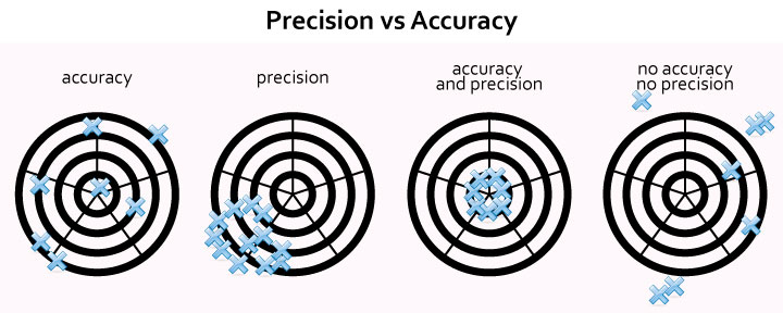
Accuracy means the data points are dispersed, but when you take the mean of those points, that mean (“sample mean”) is nearby the population mean (“true mean”). Data points are “more precise” if the dispersion across data points is smaller than some other set of data points (notice how this is a comparison and not an “absolute” statement); precision says nothing about how close the average of the data points are to the “true mean.”

Keep in mind that accuracy and precision are relative descriptors; you can’t say “so-and-so is precise”; no, you can only say “such-and-such is more precise than so-and-so” or “so-and-so is more accurate than such-and-such.” So, in this case, we can infer that NBME considers “men at the urologist” to have BUNs that are closer to each other (more clustered; more precise; less dispersed) than the BUNs of “men at mall.”

Here’s a nice image:
https://medbullets.com/images/precision-vs-accuracy.jpg

get full access to all contentbecome a member

 +4  visit this page (free120#27)
get full access to all contentbecome a member

The reason something is an “autosomal recessive” disease is because the protein encoded by the gene (of which you have two alleles, of course) does something where as long as you make SOME protein, your body should be okay.

That’s kind of vague, so take the case of Cystic Fibrosis: you don’t present with Cystic Fibrosis if you have at least one functional allele -- that’s because CFTR protein is a protein that (in the case of bronchiole tissue) moves Cl- from inside cells to the outside lumen, which brings with it H2O and keeps the bronchiole lumen nice and watery, and fluid and non-viscous and non-pluggy.

So long as you make enough of this protein, you don’t “need” both alleles to be good; the good allele can “make up for” (make enough of the protein product to compensate for) the “broken allele.” So, once again, understanding the pathophys of a disease allows you to reason through and predict things like disease penetrance and expressivity.

get full access to all contentbecome a member

 +11  visit this page (nbme22#47)
get full access to all contentbecome a member

You have to think about it this way: the basement membrane is the “scaffolding” on which [restorative] healing occurs. So, yes, stem cells (type II pneumocytes) would be involved in that healing process but they couldn’t restore the normal architecture (“no abnormalities”) without the ‘skeleton’ of the basement membrane telling them where to go, in what direction to grow, which way is “up”, etc. If the basement membrane is destroyed, you can still get healing, but it won’t be organized healing -- it’ll be disorganized healing, which does not appear as normal tissue. (Disorganized healing is better than no healing, but without a BM, the regenerating cells don’t have any “direction” and therefore can’t restore the normal architecture.)

get full access to all contentbecome a member
drdoom  by "restorative" i mean healing which restores the previous (and normal) tissue architecture. for that to happen, you need an intact basement membrane! +2
nwinkelmann  Yes, this a great summary to the post by @bubbles and the article he posted! Another way to think of the question is not, what causes repair, but what causes irreversible injury/fibrosis. That article explained an experiment that showed TGF-beta was necessary to initiate fibrosis, but if BM was intact and TGF-beta was removed, the fibrosis didn't persist, i.e. intact BM is protective against TGF-beta. https://www.ncbi.nlm.nih.gov/pmc/articles/PMC2645241/ +1

 +3  visit this page (nbme22#41)
get full access to all contentbecome a member

If I gave you a bucket of spontaneous pneumo patients -- and you reached your hand in there and pulled one out -- what scenario would be more common: In your hand you have a smoker or in your hand you have a thin male? It’s the latter.

get full access to all contentbecome a member

 +5  visit this page (nbme22#41)
get full access to all contentbecome a member

You have to think about this using the concept of CONDITIONAL PROBABILITY. Another way to ask this type of question is like this: “I show you a patient with spontaneous pneumothorax. Which other thing is most likely to be true about that person?” Or you can phrase it these ways:

  • Given a CONDITION (spontaneous pneumo), what other finding is most likely to be the case?
  • Given a pool of people with spontaneous pneumothorax, what other thing is most likely to be true about them?

In other words, of all people who end up with spontaneous pneumo, the most common other thing about them is that they are MALE & THIN.

If I gave you a bucket of spontaneous pneumo patients -- and you reached your hand in there and pulled one out -- what scenario would be more common: In your hand you have a smoker or in your hand you have a thin male? It’s the latter.

get full access to all contentbecome a member
someduck3  Is this the best approach to all of the "strongest predisposing risk factor" type questions? +
drdoom  There is a town of 1,000 men. Nine hundred of them work as lawyers. The other 100 are engineers. Tom is from this town. He rides his bike to work. In his free time, he likes solving math puzzles. He built his own computer. What is Tom's occupation most likely to be? Answer: Tom is most likely to be a lawyer! Don't let assumptions distract you from the overwhelming force of sheer probability! "Given that Tom is from this town, his most likely occupation (from the available data) = lawyer." +4
drdoom  There is a town of 1,000 spontaneous pneumo patients. Six hundred are tall, thin and male. The other 400 are something else. Two hundred of the 1,000 smoke cigarettes. The other 800 do not. What risk factor is most strongly associated with spontaneous pneumo? (Answer: Not being a smoker! ... because out of 1,000 people, the most common trait is NOT smoking [800 members].) +5
impostersyndromel1000  this is WILD! thanks guy +3
belleng  beautiful! also, i think about odds ratio vs. relative risk...odds ratio is retrospective of case-control studies to find risk factor or exposure that correlates with grater ratio of disease. relative risk is an estimation of incidence in the future when looking at different cohort studies. +
drdoom  @impostersyndrome I love me some probability and statistics. Glad my rant was useful :P +
hyperfukus  @drdoom i hate it which is why your rant was extremely useful lol i learned a ton thanks dr.doom! +1
dubywow  I caught he was thin. The only reason I didn't pick Gender and body habitus is because he was not overly tall (5'10"). I talked myself out of it because I thought the body habitus was too "normal" because he was not both thin AND tall. Got to keep telling myself to not think too hard on these. Thanks for the explanation. +1
taediggity  It isn't just that this person has Ehlers Danlos and they're more prone to spontaneous pneumo??? +2

 +27  visit this page (nbme22#34)
get full access to all contentbecome a member

Calculations for dad. The probability of the father being a carrier is 2/3 since it is known that he doesn’t have the disease. Then the probability of him passing it on to his kid is 1/2, thus:

  • Probability of dad being carrier = 2/3
  • Probability of dad passing on disease allele = 1/2

Calculations for mom. With the Hardy-Weinberg Principle, you can figure out the probability of the mother being a carrier:

q = sqrt(1/40,000) = 1/200

So, 2pq = 2 * 1/200 * 199/200, which is approx 1/100.

For the child to get the allele from mom, two things need to happen: (1) mom must be a carrier [“heterozygote”] and (2) mom must pass the allele to child:

  • Probability of mom being carrier = 1/100
  • Probability of mom passing on disease allele = 1/2

Puting it all together. Now, combine all together:

= (probability of dad being carrier) * (probability of dad passing on disease allele) * (probability of mom being carrier) * (probability of mom passing on disease allele)

= 2/3 * 1/2 * 1/100 * 1/2
= 1 in 600

get full access to all contentbecome a member
kernicterusthefrog  To quote Thorgy Thor, drag queen: "ew, Jesus, gross" +52
niboonsh  This question makes me want to vomit +14
drdoom  lol +
5thgencephalosporin  okay wow +
tekkenman101  You can make this a lot quicker by using simple rules for autosomal recessive diseases. 1) Unaffected parent with affected lineage will be a carrier 2/3 of the time (Aa) 2) frequency in population is super low so you can ignore P and just use 2(q) in order to calculate carrier frequency. So take the square root of the homozygous recessive frequency (1/40,000) and just plug it in: 2(.005) = .01 3) The odds of their child being affected with the two parents being assumed hypothetical carriers is 1/4 (aa) or .25. Dad's carrier chance (2/3) x Mom's carrier chance (.01) x child's chance of being recessive (.25) +1

 +4  visit this page (nbme22#37)
get full access to all contentbecome a member

Here’s another very nice one that superimposes the pathway onto a simplified brainstem drawing (nice for the anatomical relations):

https://webeye.ophth.uiowa.edu/eyeforum/cases-i/case252/Fig2-INO-LRG.png
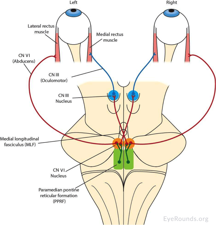
Source article:

https://webeye.ophth.uiowa.edu/eyeforum/cases/252-internuclear-ophthalmoplegia.htm

To see even more, try google image search on “medial longitudinal fasciculus”:

https://www.google.com/search?q=medial+longitudinal+fasciculus&tbm=isch

get full access to all contentbecome a member
endochondral1  what is A and B in this pic? i knew it was dorsal pons ipsilateral but i just didn't know what part that was on the pic? +1
nwinkelmann  A and B are the superior cerebellar peduncles.http://what-when-how.com/wp-content/uploads/2012/04/tmp15F2.jpg +1

 +1  visit this page (nbme22#37)
get full access to all contentbecome a member

Nice schematic of how horizontal gaze is coordinated through the abducens/MLF/oculomotor pathway:

https://n.neurology.org/content/neurology/70/17/e57/F1.large.jpg

In the diagram, the system is coordinating gaze toward pt’s left, which (conveniently) is the same as in the stem.

Source article: https://n.neurology.org/content/70/17/e57

get full access to all contentbecome a member

 +15  visit this page (nbme24#45)
get full access to all contentbecome a member

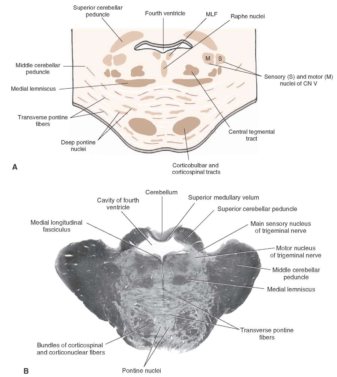
The stem is describing sequelae of posterior inferior cerebellar artery occlusion, resulting in Wallenberg syndrome. Here’s a nice schematic of the affected nuclei and brain stem regions:

https://i.ytimg.com/vi/A8S3B9p1t_g/maxresdefault.jpg

... and a 6-minute YouTube video that walks you through it:

https://www.youtube.com/watch?v=A8S3B9p1t_g

get full access to all contentbecome a member
nbme4unme  Great video! Very, very solid review of brainstem anatomy. +
suckitnbme  This image was surprisingly interpretable for NBME standards +16
aneurysmclip  and the fact that all you needed to know was the side of the lesion to answer tbh lmao, but other than that localizing to medulla wasn't hard. +6
medguru2295  Actually, they were quite nice. You didn't even have to know what side. There was no option for left medulla. +3

 +95  visit this page (nbme24#2)
get full access to all contentbecome a member

After the cuff is tied, the cells and tissue distal to the cuff will continue consuming ATP (ATP->ADP), but no fresh blood will be delivered to “clear” what will be an accumulating amount of ADP and other metabolites. ADP (=Adenosine) is itself a proxy of consumption and drives vasodilation of arteries! (Evolution is smart!) Increasing ADP/Adenosine in a “local environment” is a signal to the body that a lot of consumption is occurring there; thus, arteries and arterioles naturally dilate to increase blood flow rates and “sweep away” metabolic byproducts.

get full access to all contentbecome a member
lispectedwumbologist  You're a good man. Thank you. +1
drdoom  So glad it helped! +1
seagull  very well put, thank you +2
aisel1787  gold. thank you! +
pediculushumanus  beautiful explanation! +2
rockodude  this explanation was on par with Dr. Sattar IMO +3
flvent2120  Just to add on to this: FA2020 pg. 297. CHALK (Calcium, H+, Adenosine, Lactate, K+) is known to vasodilate muscles during exercise as well as regulate sympathetic tone of arteries at rest +3
omarjenny  Dr Sattar and pathoma failed alot people don't recommend them. +

 +7  visit this page (nbme24#25)
get full access to all contentbecome a member

EBV is not a “respiratory virus” -- it’s a B cell virus. It infects B cells; not laryngeal cells.

Even though you might associate it with the “upper respiratory tract” (=kissing disease), it doesn’t cause respiratory inflammation since that’s not its trope. B cells are its trope! That’s why EBV is implicated in Burkitt Lymphoma, hairy leukoplakia and other blood cancers. (EBV is also known as “lymphocryptovirus” -- it was originally discovered “hiding” in lymphocytes of monkeys.) So, EBV = think B cells. From the MeSH library:

The type species of LYMPHOCRYPTOVIRUS, subfamily GAMMAHERPESVIRINAE, infecting B-cells in humans. It is thought to be the causative agent of INFECTIOUS MONONUCLEOSIS and is strongly associated with oral hairy leukoplakia (LEUKOPLAKIA, HAIRY;), BURKITT LYMPHOMA; and other malignancies.

https://meshb-prev.nlm.nih.gov/record/ui?name=HERPESVIRUS%204,%20HUMAN

get full access to all contentbecome a member

 -1  visit this page (nbme20#29)
get full access to all contentbecome a member

[deleted]

get full access to all contentbecome a member

 +2  visit this page (nbme21#21)
get full access to all contentbecome a member

Also consider this great description from the NIH’s MeSH database:

INCIDENCE: The number of new cases of a given disease during a given period in a specified population. It also is used for the rate at which new events occur in a defined population. It is differentiated from PREVALENCE, which refers to all cases, new or old, in the population at a given time.

https://meshb.nlm.nih.gov/record/ui?ui=D015994

get full access to all contentbecome a member
questioneverything  The prevalence of chlamydia in this group would be 0. It is not a chronic disease. +

 +10  visit this page (nbme21#21)
get full access to all contentbecome a member

Don’t forget that incidence is the number of new cases which emerge in an unaffected population. Incidence is trying to get at the question -> “In a given year, how many new people develop this disease?”

In other words, you cannot count people who already have the disease. You have to exclude those people from your calculation. You want to know, among all the people out there who DO NOT have the disease, how many times this year was someone (newly) diagnosed?

Said differently still, you don’t want to “double-count” people who developed the disease before your study. As an epidemiologist, that would screw up your sense of how infective or transmissible a disease is. You want to know, “from time1 to time2 how many new cases emerged?”

get full access to all contentbecome a member
questioneverything  You would count the total risk pool. Chlamydia is not a chronic disease so you would treat those 500 people and they would return to the risk pool. +2
drdoom  But you would first have to determine that they CLEARED the infection. What if you gave them tx and then they come back and say, "doc i got the chlamydia" -- is this a new case or did the tx fail? You're assuming it cleared but maybe it didn't. That's why you want to EXCLUDE from the start anyone who might already have disease of interest. +7

 +20  visit this page (nbme21#21)
get full access to all contentbecome a member

2,500 students ... but you find out during your initial screen that 500 already have the disease. So, strikeout those people. That leaves 2,000 students who don’t have the disease.

Over the course of 1 year, you discover 200 students developed the infection. Thus:

200 new cases / 2,000 people who didn’t have the disease when you started your study = 10 percent

Tricky, tricky NBME ...

get full access to all contentbecome a member
sympathetikey  Ah, I see. Thank you! +
niboonsh  Im mad at how simple this question actually is +7
sahusema  Incidence is measured from those AT RISK. People with the disease are not considered to be at risk. So 2500 - 500 = 2000 people at-risk. Of those 2000, within one year 200 develop the disease. So 200/2000 of the at-risk population develop the disease. 20/2000 = 10% = incidence +3
daddyusmle  fuck im retarded +2
skonys  Must be Florida State University.... +
l0ud_minority  What if they got chlamydia again how would this change the numbers? +

 +58  visit this page (nbme21#49)
get full access to all contentbecome a member

The synthesis of virtually all proteins (mRNA->peptide) occurs in the cytoplasm.[1] That’s where all ribosomes reside, after all. Ribosomes, which are mostly just rRNA (~2/3 rRNA + 1/3 protein*, by weight), are assembled in the nucleus but only do their stuff once they get to the cytoplasm.

For a protein to leave its original hometown of the cytosol and become a resident of the nucleus or, say, the endoplasmic reticulum, it needs to have a little string of amino acids which shout “I belong in the nucleus!” or “I belong in the endoplasmic reticulum!”

Proteins ultimately destined for the ER contain an unimaginatively named string of amino acids known as “signal sequence,” which, for the purposes of the Step 1, is always at the N-terminus. The signal sequence tells other cytosolic proteins, “Hey! Take me (and the rest of the peptide of which I am part) to the ER!”

In the absence of this signal, a protein will remain in its “default” home of the cytosol.

Here’s a nice schematic showing the flow of proteins from initial synthesis to final destinations:


Endnotes

  1. “The synthesis of virtually all proteins in the cell begins on ribosomes in the cytosol.” (Essential Cell Biology, Alberts et al., 2014, p. 492)

*If you really want your mind blown, consider that even the protein subunits that make up that 1/3 of a ribosome are themselves initially synthesized in the cytosol; later, they are transported back into the nucleus via the nuclear pore.

get full access to all contentbecome a member
qball  Awesome explanation. Now explain it to me like I'm 5. +13
drdoom  All baby peptides are born in the cytosol. But some baby peptides have a birthmark at their N-terminus. The birthmark tells a special mailman that this baby needs to be delivered somewhere else. If you chop off the birthmark — or erase it somehow — the mailman never knows to take baby to its true home. The end. Now go to sleep or Santa won’t bring your presents. +83

 +0  visit this page (nbme21#28)
get full access to all contentbecome a member

Here’s one way to process-of-eliminate “decreased hydrogen-bond formation”: I’m not a big fan of this line of reasoning, but technically alanine as a side group has more hydrogens* for potential hydrogen bonding than glycine:

alanine: —CH3
glycine: —H

So, “technically,” alanine would permit more hydrogen-bond formation, which might allow you to eliminate that choice.

That said, it seems almost impossible to rule out (without very technical knowledge or some provided experimental data) that the slightly larger alanine does not impair hydrogen bonding between collagen molecules via steric (spatial) interference. In simpler terms, since alanine is larger, you would think that it must somehow interfere with the hydrogen-bonding that occurs with the wild-type glycine.

---
*Strictly speaking, it’s not the number of hydrogens but also the strength of the dipole that facilitates hydrogen bonding: a hydrogen bound to a strongly electronegative molecule like fluorine will “appear” more positive and, thus, hydrogen-bond more strongly with a nearby oxygen (compared with a hydrogen connected to carbon, for example).

Further reading:

  1. https://www.chem.purdue.edu/gchelp/liquids/hbond.html
get full access to all contentbecome a member
hungrybox  Appreciate the effort but this is far too long to be useful. +30
drachenx  hungrybox is a freaking hater +
drdoom  @drachenx haha, nah, coming back to this i realize i was probably over-geeking lol +1
blueberrymuffinbabey  isn't the hydrogen bonding dependent on the hydroxylated proline and lysine? so that wouldn't really be the issue here since those aren't the aas being altered? +
drdoom  @blueberry According to Alberts’ MBoC (see Tangents at right), hydroxylysine and hydroxyproline contribute hydrogen bonds that form between the chains (“interchain”, as opposed to intra-chain; the chains, of course, are separate polypeptides; that is, separate collagen proteins; and interactions between separate chains [separate polypeptides] is what we call “quaternary structure”; see Tangent above). And in this case, as you point out, the stem describes a Gly->Ala substitution. That seems to mean two things: (1) the three separate collagen polypeptides will not “pack [as] tightly” to form the triple helix (=quaternary structure) we all know and love and (2) proline rings will fail to layer quite as snugly, compromising the helical conformation that defines an alpha chain (=secondary structure; the shapes that form within a single polypeptide). +
tadki38097  also you can't H bond with carbon, it's not polar enough +
amy  FA2020 P50 state: formation of procollagen(which is the triple helix structure) via hydrogen and disulfide. So A is incorrect bc there are no collagen molecules yet (2nd structure happens at procollagen level) +
drdoom  @amy triple helix is a quaternary structure (since triple helix is a shape that forms BETWEEN separate or “adult” pro-collagen/collagen peptides). Primary, secondary and tertiary structures are descriptions of a single polypeptide. Once you have 2 or more polypeptides interacting with each other (e.g., Hemoglobin molecule) you have quaternary structure. +

 +8  visit this page (nbme21#23)
get full access to all contentbecome a member

Vasoconstriction (narrowing of a tube) will cause the flow rate to increase through that tube, which decreases radial/outward pressure. The faster a fluid moves through a tube, the less “outward” force it exerts. (This is known as the Venturi effect.)

get full access to all contentbecome a member
hungrybox  not seeing how this is relevant +9
sympathetikey  He's showing how A & B are incorrect @hungrybox +7
nerdstewiegriffin  what a moron @hungrybox is !! +2
leaf_house  MCAT flashbacks on this image +1

 +11  visit this page (nbme20#6)
get full access to all contentbecome a member

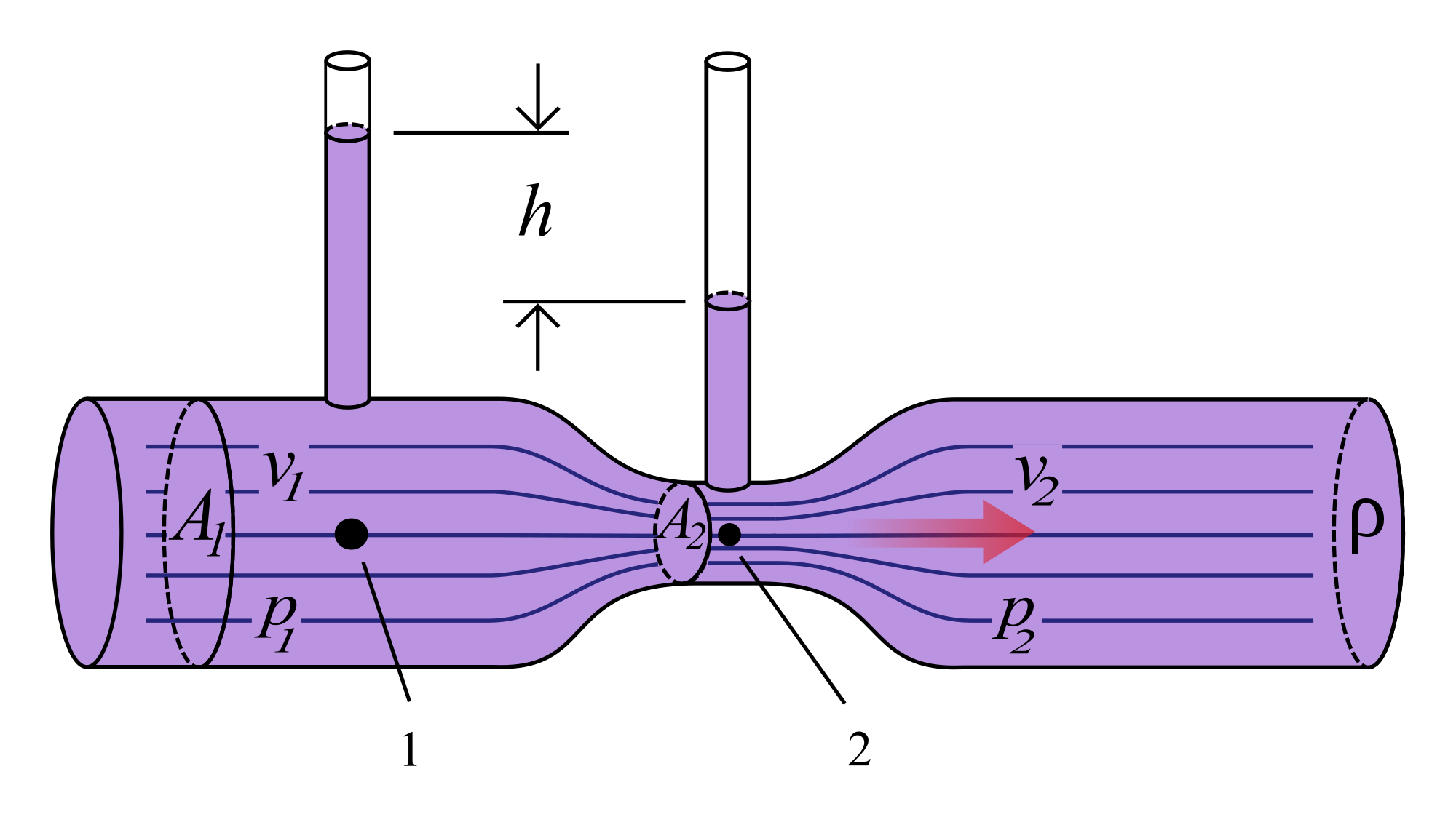
This is an interesting one. I like to remember it this way: in people with narcolepsy, all the “right kinds” of sleep are happening at all the “wrong times” of day. During the day, when a power nap would typically throw you immediately into REM, this kid is only entering Stage 1 or 2 (lightest sleep = slightest noises jar him back to reality). At night, when he should peacefully drift into Stage 1, 2, and so on, he instead completely zonks out. Classic narcolepsy.

From UpToDate: “Narcolepsy can be conceptualized as a disorder of sleep-wake control in which elements of sleep intrude into wakefulness and elements of wakefulness intrude into sleep.”

get full access to all contentbecome a member
chextra  Isn't REM a rather light sleep stage? Brain waves during REM are very similar to awake states. I think you even wake up briefly in the middle of REM sleep. I don't think FA gave me a great understanding of narcolepsy, but I see it as going from awake to REM (light) for any kind of sleep, daytime or night time. +
sammyj98  I'm definitely not ace on this subject, but I think the brain waves present in REM are similar to wakefulness because of the dreaming component. I think of it as though the brain has to go through a process of hypnotizing the body into a state of relaxation, and then properly paralyzing it, and then it can simulate wakefulness (dreaming) to go through with it's defragging of the hard drive. So REM is actually the deepest sleep because the body is fully paralyzed. Please someone correct me, this is probably an inacurrate perspective. +4
pg32  FA says that narcolepsy has nocturnal AND NARCOLEPTIC sleep episodes that start with REM sleep... So is @drdoom correct? FA seems to disagree regarding the daytime sleep pattern. +8

 +1  visit this page (nbme20#15)
get full access to all contentbecome a member

The more general principle: endothelia vasodilate in the presence of high CO2; you gotta get rid of that acid somehow! Can’t let it accumulate, as lower pH within a “micro-environment” affects structure/efficiency of enzymes, proteins, etc. The more acidic a local environment, the more you expect nearby vasculature to dilate (as a means of increasing flow rate, thereby ferrying off accumulate acid).

The anesthesiologist can exploit this mechanism. By hyperventilating (blowing off CO2), the brain vasculature senses a low CO2 / “hunky-dory state,” which requires no vasodilation. In other words, the vasculature does not need to continue the ATP-consuming practice of synthesizing Nitric Oxide (NO).

get full access to all contentbecome a member
hello  But, the Q-stem states the anesthesiologist is HYPOventilating the patient. +4
drdoom  decreasing respiratory rate = retention of CO2 = vasodilation of brain arteries = more filling of tubes = greater intra-cranial pressure +2
drdoom  @hello shoot, you're right! i ended my explanation with the example of HYPERventilation when i should have done the opposite! (sorry!) ... edit: "By HYPOventilating (retaining CO2), the brain vasculature senses a high CO2 environment and vasodilates = increases intra-cranial filling and pressure!" +3
dulxy071  @drdoom could you please elaborate on your point. +

 +4  visit this page (nbme19#13)
get full access to all contentbecome a member

This is essentially a formal logic question. Logically speaking, the question asks us to identify a mechanism that tumor suppressors have which proto-oncogenes do not. In other words, what is a mechanism shared by all known tumor suppressors but not shared by any known proto-oncogenes? For that reason, it can’t be phosphorylation; sure, phosphorylation is a mechanism of tumor suppressors but it’s also a mechanism of many known proto-oncogenes.

get full access to all contentbecome a member

 +11  visit this page (nbme19#4)
get full access to all contentbecome a member

Inability to maintain an erection = erectile dysfunction. So now the question is "Why?"

Fatigue, difficulty sleeping, difficulty concentrating is starting to sound like depression. "Difficulty concentrating" might be interpreted as impaired executive function or the beginnings of vascular-related dementia (dementia related to small but numerous cerebral infarcts), but on Step 1 dementia will be blatant (i.e., "lost his way home," "wandering," etc.).

Depression is actually common after a debilitating event like stroke, as you might expect. With depression comes a loss of sexual interest and desire—that is decreased libido.

One can make the argument that a "vascular patient" might have some issues with his "pipes" (arteriosclerosis, parasympathetic/sympathetic dysfunction) and, for this reason, nocturnal erection should be decreased; but note that nothing is mentioned about long-standing vascular disease (no hx of hypertension).

As a result, the best answer choice here is C. (Libido decreased but nocturnal erections normal.) The big question I have is, how the heck does this guy know he's hard when he's asleep!!? :p

get full access to all contentbecome a member
cbay0509  thank you +1
ilikedmyfirstusername  there are several UWorld questions about psychogenic ED with the answer being normal libido and normal nocturnal erections, idgi +15
djeffs1  Yeah NBME says its C, but I still think with a recent stroke you can't bank on normal nocturnal erections... +
drdoom  @djeffs nocturnal erections happen at the level of the spinal cord (S2–S4)! a “brain stroke” (UMN damage or “cortical damage”) would not kill your ability to have nocturnal erections! https://en.wikipedia.org/wiki/Nocturnal_penile_tumescence#Mechanism +2
drjo  fatigue, difficulty sleeping and concentrating could be depression or hypothyroidism both of which can cause decreased libido +
jurrutia  @djeffs1 when you say NBME say's it's C, how do you know that's the official answer? Did NBME post the answers somewhere? +
djeffs1  in the versions I purchased from them they highlight the correct answer in the test review +1
shieldmaiden  For me the keyword in the stem is "maintain"; he can maintain an erection, therefore nocturnal erections must be normal. Libido, on the other hand, is psychologically driven, so if he is depressed (trouble sleeping, concentrating, fatigue, recent major health problem) then the strength towards any kind of desire, including sexual, will be low +2
chaosawaits  His nocturnal erections are normal because his spinal cord is not damaged. His libido has decreased because he's showing signs of depression. +


Subcomments ...

submitted by azibird(279), visit this page
get full access to all contentbecome a member

The only reliable way to differentiate between PCP and cocaine on these exams: + nystagmus (not present here) + catatonia/sedation

Weird facts about PCP intoxication: + May wax and wane between extreme agitation and sedation + Blank stare and flat affect + Rigidity + May not have hypertension or tachycardia

get full access to all contentbecome a member
azibird  Formatting :( +
drdoom  Start lists on new lines; maybe also leave a blank line between the list and preceding paragraph +


submitted by azibird(279), visit this page
get full access to all contentbecome a member

I am NOT giving a stress test to every 50 year old male smoker with hypertension and family history of MI who wants to exercise. That would be so many patients and seems like a bad use of resources.

Tell me why I'm wrong. Is it because he has so many risk factors for CHD (age, sex, smoker, hypertension, family history)?

From UTD: "CHD screening tests are generally not recommended for asymptomatic patients, and cardiac stress testing should not be part of a routine annual physical or health screening examination. However, there are other patients in whom we perform stress testing: patients who need reassurance that it is safe for them to be active, in which a stress test can help providers delineate an activity program with specific levels of exercise to achieve; and patients who have an occupation in which high levels of exertion may be routinely required (eg, farmer)." https://www.uptodate.com/contents/screening-for-coronary-heart-disease

"Rare exceptions are patients with multiple risk factors" https://www.uptodate.com/contents/selecting-the-optimal-cardiac-stress-test

get full access to all contentbecome a member
drdoom  Your reasoning makes sense to me. UTD >> NBME +


submitted by bfinard1(3), visit this page
get full access to all contentbecome a member

Can someone explain why cardiac output is high in septic shock?

get full access to all contentbecome a member
drdoom  Bacteria and bacterial components (like LPS) in circulation trigger massive release of cytokines (Interleukin-1, Interleukin-6 and TNFa), resulting vasodilation. Heart rate increases to maintain decent BP. +1
bfinard1  Is stroke volume not affected by that massive vasodilation? I would think if venous system is vasodilated then you'd have reduced EDV from reduced blood flow to heart +
zedora  Both of you are correct. In septic shock there is a massive vasodilation. In order to compensate for the reduced blood pressure, there is an increased heart rate. Now, keeping this in mind, what is the CO formula? CO = HR x SV Right? Lets say under normal conditions HR is 60 & Stroke volume is 50. Your cardiac output = 60 x 50 = 3000. Now in septic shock, your heart rate is massivly increased but your stroke volume is decreased minimally. So lets plug in the numbers. Lets say, under septic shock, HR = 150 & stroke volume is now 30. The cardiac output is now gonna be = 150 x 30 = 4500, hence your CO is increased. In Septic Shock, the heart rate is massively increased compared to the amount of SV decreased. +
drdoom  @bfinard1 By vasodilation, I almost exclusively mean arterial vasodilation. When it comes to CV, I always work backward from “first principles”, and in my view the first principle of the CV system is, “Maintain pressures to maintain good flow.” All other accommodations of the CV system (changes in inotropy[strength], chronotropy[time], vasoconstriction and vasodilation) are in *service* to maintaining flows. Without good flows, you get the thing human tissues like the least: not lack of oxygen but accumulation of CO2 (and the acidity that goes with it). +
drdoom  So, all that was a long-winded way of saying that Cardiac Output will remain high when the body is producing higher-than-desired levels of CO2 (when organs and the immune system have gone into overdrive to respond to a threat or to address decompensation in some other part of the system); the plummeting of Cardiac Output heralds the beginning of the end. It signifies that the stresses being imposed on the body exceed the capabilities of the system. +


submitted by bwdc(697), visit this page
get full access to all contentbecome a member

Malignant pleural effusion. Cancer is full of protein.

get full access to all contentbecome a member
nbmehelp  ??????????? +
drdoom  hi @nbmehelp. if you are confused about a comment or unhappy with an explanation, please pose a specific question so we can help answer or clarify. thanks! +
rthavranek  But malignancy effusion also has glucose < 60 mg/dL so I was between that and increased protein. My reasoning was that cancer cells have high metabolic rate so they require increased energy/glucose +1
cbreland  I thought the same thing @rthavranek , all exudative effusions should have increased protein. That being said, still picked decreased glucose +


submitted by agurl1000(5), visit this page
get full access to all contentbecome a member

Could someone explain to my why it is Factor X? Like I understand that PTT and PT are both elevated. I picked Factor X and then changed it to "hydrolysis of.." because I thought with a Factor X deficiency, wouldn't you have a prolonged thrombin time? I might be making this more complicated, but doesn't Factor X play a role in thrombin activation, so lack of it would lead to a prolonged thrombin time?

get full access to all contentbecome a member
drdoom  a person with FXD (factor x deficiency) will have "prolonged thrombin time" inside their body (in vivo), yes, but their THROMBIN_TIME (the lab test) will be completely normal +1
drdoom  when they run the lab test, what they do is just add a bunch of lab-grown (recombinant?) ACTIVATED thrombin to your plasma and then count the seconds until a clot forms. that’s your THROMBIN_TIME. notice how if you have fucked up Factor X, it doesn't matter because the lab guy is adding NORMAL, lab-grown/off-the-shelf activated thrombin to your plasma sample. +1
drdoom  in other words, by adding the lab thrombin, we "circumvent" any problems leading up to AND INCLUDING the production of functional thrombin. (since perfectly functional thrombin is what’s being supplied by the lab guy.) THUS what THROMBIN_TIME (the test) is really asking is, “Is there anything inside this patient that is interfering with the conversion of fibrinogen->fibrin?” +1
drdoom  SO, what the test should REALLY be called is not THROMBIN_TIME (which is confusing as hell) but TIME_REQUIRED_TO_MAKE_FIBRIN_FROM_FIBRINOGEN +1
agurl1000  THANK YOU SO MUCH! THIS MAKES SO MUCH SENSE! +
drdoom  ☺️☺️~ +
drdoom  p.s. I did a little more reading and it seems like thrombin is derived from plasma donors (not made recombinantly in a dish😁 sorry!) +


submitted by medstruggle(21), visit this page
get full access to all contentbecome a member

Why is it aphthous ulcers if there are no GI symptoms? Why can’t it be herpes zoster?

get full access to all contentbecome a member
colonelred_  It’s just canker sores, they come and go. I think in herpes the gingivostomatitis really only happens when you first get infected. After that you just get recurrent cold sores. +4
hyoid  Herpes zoster is not the same as herpes simplex virus. +31
bigjimbo  you would see dermatome rash in zoster +3
kateinwonderland  cf) Just in case someone wanted to know the causative organism of aphthous ulcers :The precise cause of canker sores remains unclear, though researchers suspect that a combination of factors contributes to outbreaks, even in the same person. Unlike cold sores, canker sores are not associated with herpes virus infections. +9
charcot_bouchard  Herpes Zoster doesnt cause gingivostomatitis. Herpengina can cause vesicular lesion in mouth but happens to children in summer season by entero virus +
drdeeznuts1  I'm wondering if this could be a mild case of Behcet syndrome without genital involvement +
sherry  It sure can be Behcet or Pemphigus if the q provides us with more info. Canker sores just come and go for years with unclear mechanism. Also herpes zoster is shingles by VZV, not HSV1. +2
avocadotoast  Most pictures on google show herpangina being present on the hard palate/throat, while aphthous ulcers are commonly on the lower lip. I think his lack of genital lesions are pointing us away from herpangina. +
drdoom  Anyone still curious about the pathophys of aphthous ulcers can check out the thread above: https://nbmeanswers.com/exam/nbme24/964#4853 +


submitted by thisshouldbefree(51), visit this page
get full access to all contentbecome a member

I get these VERY often. I seem to get them only after eating something that can damage the mucosa in my mouth such as hard chips (Tostitos, very jagged very pain). These are NOT cold sores (HSV). This is simply a aphthous ulcer; they can be stress induced (studying for hard test). they are very painful and do reoccur. there are no associated sx with them.

get full access to all contentbecome a member
j44n  they're also caused by stress. I got 3 in my mouth the day after Prometric canceled my test for the 3rd time! +15
ih8payingfordis  In layman terms, these things are called canker sores. I also get them on my tongue sometimes - super painful especially when it makes contact with anything salty. Just like @thisshouldbefree, it's usually preceded by mucosa damage (ie biting my tongue accidentally while eating) +2
xmen  test in 2 weeks. i have them now +1
drdoom  The physiological corollary of psychological “stress” is cortisol and its many cousins (corticosteroids, et cetera). Cortisol suppresses the “normal” but energetically costly process of dermal maintenance and growth (basal stem cell division, skin + epidermal turnover, including in the mucosa). So, in the presence of high “stress”, normal mucosal maintenance is sacrificed. This is because some other, more important existential threat is happening in the body. The result is stuff like aphthous ulcers, since your body is literally neglecting your mucosa, allowing it to “thin out” to preserve energy to address some other major systemic issue. +1
drdoom  From https://www.ncbi.nlm.nih.gov/pmc/articles/PMC5181654/?report=classic: The glucocorticoid receptor (GR) is highly expressed in the basal cells but it is barely detectable in the other layers of the epidermis (22). The localization of the GR in basal corneocytes, as well as the negative effects of glucocorticoids on keratinocyte growth factor and type-I and-III collagen gene expression (23–26) suggest that endogenous cortisol in CS suppresses not only wound healing but also normal skin growth and turnover. +
drdoom  ^ btw, CS = Cushing Syndrome (hypercortisolism) +
jbrito718  Herpangia would occur in the posterior oropharynx--> caused by Coxsakie A NOT HSV +
meryen13  I have some in my mouth rn... 2 weeks to my step... they can be food or stress induced and its more common in some regions more than others. in Middle East and Asia it seems to be common. +


submitted by pingra(6), visit this page
get full access to all contentbecome a member

I thought of this as a giant physiologic shunt (ie, due to the pneumothorax there is no ventilation to an entire lung, as a consequence you retain CO2) - not sure if this is the actual mechanism but it helped me get this question right

hopefully this helps!

get full access to all contentbecome a member
drdoom  this definitely makes sense to me, especially if it happens “acutely”/suddenly. if someone gets a lung or lobe removed, e.g., cancer, my guess is that the reminaing lung would “remodel” over time and recoup at least some of that lost surface area — in the same way new anastomoses form in the weeks or months after near-complete artery blockage (as guided by VEGF elaboration) +
drdoom  but in the case where it happens “all of sudden”, i totally agree you’re going to get CO2 retention +


submitted by sugaplum(487), visit this page
get full access to all contentbecome a member

These are symptoms of acute benzo withdrawl

get full access to all contentbecome a member
drdoom  holy cow you are crushing it right now. frickin POWER SESSION +3


submitted by beetbox(6), visit this page
get full access to all contentbecome a member

Can someone explain more on how to tackle these types of questions? I suck at these questions for real... To me, he sounded pretty sane and reasonable (does not wish to waste other people's money). Sure he might be under slight depression judging how he has a terminal illness and his statement on how nobody cares for him. But unless he is incoherent or displaying magical thinking, signs of loss of memory etc., why should he be evaluated on decision-making capacity?

get full access to all contentbecome a member
drdoom  in medical parlance, you can be so depressed that you're actually cognitively impaired. this is known as pseudodementia. thus, you need to figure out: “is this guy so depressed we can deem him incompetent to make decisions?” +
rockodude  he says he has an invention to cure arthritis in 6 months he'll be back.. not normal imo. at least for this question thats the line that made me think does this person have capacity +


submitted by keycompany(351), visit this page
get full access to all contentbecome a member

Flow Rate = Velocity x Cross-Sectional Area

2 cm^2 x 20 cm/sec x 60 sec/min x 1 L/1,000 cm^3 = 2.4 L/min

1,000 cm^3 = 1 L

get full access to all contentbecome a member
seagull  Well, I missed this one. I don't even feel bad. +78
link981  @keycompany a small typo, 100 cm^3 = 1 L not 1000cm^3. 1000 mL^3= 1 L +1
hello  @keycompany how did you edit your original comment to fix your typo? +1
winelover777  Pretty sure @keycompany was correct. 1 L = 1000 cm^3. Otherwise the answer would be 24. +4
drdoom  1 centimeter is a distance. (A line.) +1
drdoom  If we multiply a line by another line, we get a surface area. (A piece of paper.) +1
drdoom  If we multiply the piece of paper by another line, we get volume. (A cube. A box.) +1
drdoom  If we fill the box with a fluid, we will have 1 mL of this fluid. +1
drdoom  If we have a thousand of these boxes, we have 1 L of fluid. +1
drdoom  1,ooo mL = 1 Liter = 1,ooo centimeter³ +1


submitted by johnthurtjr(168), visit this page
get full access to all contentbecome a member

here's a google

get full access to all contentbecome a member
johnthurtjr  FTR I had no idea this was a thing, and was pretty disappointed in myself when the google search had it in big bold letters right in my face. +3
drdoom  via @johnthurtjr link: "Testosterone and other androgens have an erythropoietic stimulating effect that can cause polycythemia, which manifests as an increase in hemoglobin, hematocrit, or red blood cell count." https://www.medscape.com/viewarticle/773465 +3
meningitis  I guess that's another reason for steroids and doping up. +8
drschmoctor  For once I feel like I've been led astray by Pathoma. My instinct was to go with hemoglobin, but I talked myself out of it after remembering Dr. Sattar saying that the reason why women have lower hemoglobin is due to menstruation. +2
fexx  F U testosterone! and F U NBME 22 question +1
schep  I only knew this because there are three (at least three, maybe more that I don't know) contraindications to giving testosterone replacement therapy: +OSA +prostate cancer +hematocrit >50% +2
kayla  @drschmoctor; I still think it lines up with the correct reasoning; during the menstrual phase ( in addition to loosing hemoglobin in the blood) there are also very low levels of the androgen hormones that usually serve as a stimulating effect on hemoglobin... +
abhishek021196  Fortunately, we were taught this in med school - testosterone stimulates erythropoiesis by stimulating EPO and recalibrating the set point of EPO in relation to hemoglobin and by increasing iron utilization for erythropoiesis. https://www.ncbi.nlm.nih.gov/pmc/articles/PMC4022090/ +


submitted by syoung07(58), visit this page
get full access to all contentbecome a member

if you are 68 and still dating Palmala Handerson, your libido isnt low my friends.

get full access to all contentbecome a member
drdoom  amen +


submitted by cbreland(195), visit this page
get full access to all contentbecome a member

If you knew that basement membrane disruption prevents restoration of normal tissue (repeat from another NBME), then you missed this because you didn't know what "preclude" means.

Missing questions I miss due to lack of vocab and grammar, you love to see it

get full access to all contentbecome a member
cbreland  Say a prayer for me +3
drdoom  you can include. you can exclude. or you can altogether preclude. +2
drdoom  all these words, along with “claudication”, share the same Latin root: clud = claud (to shut) +1
dhpainte22  Missed it for the same reason :( +1
hamdiabdeen  Can someone explain why basement membrane is correct answer as opposed to capillaries or Type I pneumocytes? +
hamdiabdeen  You right, thank you +
randi  guess I gotta make an Anki card for the definition of preclude then +
sparta  does anybody have a dictionary Anki deck? +1
notpennysboat  Guilty. I spent good time on this question trying to figure it out, and still got it wrong +


submitted by haozhier(23), visit this page
get full access to all contentbecome a member

Can someone please explain to me: If the posterior 1/3 of the tongue is developed from 3rd and 4th pharyngeal arches, why is it wrong to choose pharyngeal arch?

get full access to all contentbecome a member
therealslimshady  Welcome to NBME +2
mutteringly  First time? (meme) +4
drdoom  That would be like choosing “blastula” if it were an option: it's not wrong but there's a more precise answer. +
pontiacfever  That is wrong. They're indicating towards thyroglossal duct/thyroid which originates from Pharyngeal pouch not arch. secondly, they're asking that the mass originated from which structure. So, as we know it is associated to foramen cecum which is related to tongue. +
drdoom  @pontiacfever, i believe you’re responding to @haozhier’s original comment, yes? +
pontiacfever  @drdoom, yes the original comment by @haozhier +1


submitted by j44n(141), visit this page
get full access to all contentbecome a member

Just another piss poor government institution cutting corners. If you've done NBME 18 and seen the cell diagram figure it is the literal pinnacle

get full access to all contentbecome a member
drdoom  the NBME is a private, for-profit corporation. individual U.S. states use its products (i.e., the certification it gives you when you pass their exams) to determine your eligibility to practice in their state. but they are not a government entity. +1


submitted by andro(269), visit this page
get full access to all contentbecome a member

Graft Vs Host disease

Look out for Skin involvement - maculopapular rash
Enteric involvement - diarrhea and or cramping , abdominal pain .. nausea/vomiting
Hepatic dysfunction - jaundice

*** The skin , liver and intestines are the most involved affected organs

get full access to all contentbecome a member
drdoom  basic science of GVHD https://youtu.be/he2vfNZDfbY?t=522 +7
bingcentipede  Graft vs. Host disease - type IV hypersensitivity response, but this is the only one where the graft (T cells) are attacking the recipient (cells). Additionally, GvH dz is very common (at least in questions) in BONE MARROW TRANSPLANTS (also liver, but BMTs seems to pop up a lot) +2
fatboyslim  ^They are more common in bone marrow transplants because there are more T cells in the bone marrow that are transplanted from the donor to the host, hence a higher likelihood of T cells to attack the host +


submitted by tiredofstudying(71), visit this page
get full access to all contentbecome a member

FA 2020: 463

Causes of avascular necrosis: CASTS Bend LEGS. +Corticosteroids +Alcoholism +Sickle cell disease +Trauma +SLE

+“the Bends” (caisson/decompression disease) +LEgg-Calvé- Perthes disease (idiopathic) +Gaucher disease +Slipped capital femoral epiphysis

get full access to all contentbecome a member
drdoom  for bulleted lists, be sure to follow the plus sign with a space! :) +3


submitted by vshummy(184), visit this page
get full access to all contentbecome a member

I think more generally, protein folding happens at the RER and the stem says the protein doesn’t fold properly. Specifically, the most common CF mutation is a misfolded protein and the protein is retained in the RER and not transported to the cell membrane - FA 2019 pg 60.

get full access to all contentbecome a member
uroosisyed5  Which makes sense if we think about the pathophys of elevated Cl- and Na intracellularly. Both of these ions go up inside the cells due to the retention of the misfolded proteins in the RER. +
lilyo  I actually disagree with this reasoning. The pathophysiology in CFTR is not due to accumulation of misfolded proteins. It is due to decreased/absent ATP gated transmembrane Chloride channel. According to Uworld, the miscoded protein is detected by the Endoplasmic Reticulum. The abnormal protein is targeted for destruction by the proteasome and never reaches the cell surface. There is NO retention of misfolded protein, there is degradation of misfolded protein and therefore absence of chloride channels on the membrane. This is what leads to impaired removal of salt from the sweat as well as decreased NaCl in mucus. I dont think the answer should be ER. Can anyone tell me if I am missing something here that makes the answer ER as opposed to cytoplasm? Because the way I see if is misfolded proteins go form the ER into the cytoplasm to reach the proteasome and then be destructed. Uworld questions ID are 805, 802, 1514, and 1939. +19
drdoom  @lilyo The CFTR is a transmembrane protein. Like all proteins, its translation begins in the cytosol; that said, CFTR contains an N-terminus “signal sequence”, which means as it is being translated, it (and the ribosome making it!) will be transported to the Endoplasmic Reticulum.^footnote! As it gets translated, its hydrophobic motifs will emerge, which embeds the CFTR protein within the phospholipid bilayer of the ER itself! That means the protein will never again be found “in the cytosol” because it gets threaded through the bilayer (which is, in fact, how all transmembrane proteins become transmembrane proteins at the cell surface -- they have to be made into transmembrane proteins in the ER first!). +11
drdoom  @lilyo (continued) So, yes, ultimately, these misfolded proteins will be directed toward a proteasome for degradation/recycling, but that will happen as a little vesicle (or “liposome”); the misfolded protein, in this case, is not water-soluble (since, by definition, it has hydrophobic motifs which get “threaded through” a bilayer to create the transmembrane pattern), which means you won’t find it in the cytosol. +6
drdoom  ^footnote! : The movement of active* ribosomes from the cytosol to the Endoplasmic Reticulum is why we call that area of ER “rough Endoplasmic Reticulum (rER)”; on electron microscopy, that section was bespeckled with little dots; later, we (humans) discovered that these dots were ribosomes! +1
drdoom  * By “active ribosomes”, I just mean ribosomes in the process of converting mRNA to protein! (What we call “translation” ;) +1
wrongcareer69  How many goddamn ways are they going to test us on CF. I'm so over this! +2
furqanka  also in FA, under alpha 1 antitrypsin, its says 'Misfolded gene product protein aggregates in hepatocellular ER". might be the same concept. +9
joanmanuel26  According to Kaplan; All proteins that are synthesized in the ER must fold correctly in order to be transported to the golgi aparatus and then to their final destinations. If the mutation cause a misfolded protein, the result will be the loss of the protein function and, in some cases, accumulation of the protein in the ER. +1
drdoom  @lilyo Thinking about this more. You will not find the (misfolded) protein in the cytosol. The misfolded protein may be inside a proteasome—and a proteasome may live in the cytosol—but the misfolded protein itself will never appear in the cytosol. The products of its degradation might (constituent amino acids or small peptides) but if you had an antibody for the misfolded protein and asked it “Where is the misfolded protein?”, the antibody would answer: “Most of what I could find appears to be in the rER.” +
yesa  Check out the calnexin cycle, usually proteins that are made in the RER and misfolded get tagged and 'refolded' and then scanned again for proper folding...and if they really can't be refolded, they're degraded then and there! So the only place misfolded CFTR could accumulate if misfolded is....RER... (Link to calnexin cycle: https://www.google.com/search?q=uggt+in+rer&sxsrf=ALeKk02X8jH8eveQ2IVM9IsVOlO47Qjh5A:1597110732938&source=lnms&tbm=isch&sa=X&ved=2ahUKEwiI75yPhZLrAhUTZjUKHWxgCaIQ_AUoBHoECAwQBg&biw=886&bih=1045#imgrc=4UYRaaYQDDFbDM) +
amy  FA2020 47: " Absent or dysfunctional SRP (signal recognition particle) results in accumulation of protein in the cytosol" +
drdoom  @amy, that’s a good point but it assumes the protein is otherwise normal, i.e., not misfolded or “abnormal” in some other way +


submitted by imnotarobotbut(184), visit this page
get full access to all contentbecome a member

A medical student shouldn't be the one giving someone a cancer diagnosis. This is a really sensitive issue and the results should be given by someone with higher authority like a resident or attending. At the same time, you shouldn't lie to the patient and say that the results aren't back yet if they are. Best thing to do is deflect the conversation and follow up with the resident..

get full access to all contentbecome a member
drdoom  It isn’t so much “someone with higher authority” as it is someone with a license! Without a license, an individual is not permitted legally to provide clinical interpretations, as that would constitute the (unlawful) practice of medicine! +22
veryhungrycaterpillar  Laughable. 21 upvotes? The legality isn't the issue at all. If you wanna get legal: the student is protected under their preceptor's (resident's) license, as the resident is protected under their attending's, so legality isn't really the issue here. The issue truly is that someone trained in cancer counseling (which involves breaking the news in an appropriate manner) should be the one to break the news. Furthermore, once you tell the patient it is a "carcinoma", the patient is almost certainly going to want to know more details, prognosis, and discuss the management plan-- none of which the resident or the medical student are trained to provide, that's why they're waiting for a specialist (ie the oncologist) and not a filthy internist hospitalist. @imnotarobotbut was correct. +1
drdoom  Not sure what you're referring to here; a license to practice does not extend to your trainees. It's like you're suggesting if I have a license, I can let my 8th grader deliver the news or administer a test because .. they're under my supervision? What authority or rights does any medical student have? You might be confusing liability coverage with license. +
skonys  I practice dentistry under my professor's license everyday. I also give diagnoses under their license as well. I'm pretty sure that it's just that as you wont be rendering care you should defer to the person who will be treating them ie: a resident or physician. It has nothing to do with medical liability, it's just clearly above your pay grade as a student. +
drdoom  @skonys I believe your pay grade, as a student, is zero. Or more accurately, negative. Since you’re paying to be there. You’re not practicing “under your professor’s license”. You’re practicing under their supervision. Which is why you have zero authority to practice OUTSIDE their supervision. Because you are not permitted to practice +
drdoom  also @skonys: whatever it is you’re doing, it isn’t the lawful practice of anything. You are not a practitioner because you do not hold a license. “You” do not give diagnoses; you might tell a patient what you think, but as a student nothing you say would ever be recognized, legally, as a diagnosis. +


submitted by meningitis(644), visit this page
get full access to all contentbecome a member

Tanner stages start at TEN years old

Stage I:

  • I is flat, as in flat chest;
  • I is alone, as in no sexual hairs.

Stage II (2): stage II starts at 11 y/o (II look like 11)

  • 2 balls (testicular enlargement)
  • 2 hairs (pubic hairs now appearing)
  • 2 breast buds form

Stage III (3): starts at 13 y/o

  • If you rotate 3, it looks like small breasts (Breast mounds form);
  • If you squiggle the III they look like curly+coarse pubic hair
  • Increased penis length and size can be represented by: II --> III
    (your penis was thin II but now its thicker III)

Stage IV (4): starts at 14 y/o

  • First imagine: The I in IV represents the thigh, and the V in IV looks like the mons pubis between your legs:
    MEANING: you have hair in mons pubis (V) but you have a border detaining the hair from growing into thighs.
  • The V is pointy, as in now the breasts are pointy (raised areola or mound on mound)

Stage V (5): 15 y/o

  • V has no borders detaining hair from growing into thighs (pubic hair + thigh hair)
  • 5 fingers(as in hands) flattening the areolas when grabbing them (areola flatten at this stage and no more "mound on mound")

get full access to all contentbecome a member
meningitis  Sorry about the format, it came out wrong but I hope his helps. +2
drdoom  looks good to me! +25
gh889  According to FA2019, stage 2 ends at 11, stage 3 starts 11.5-13, and stage 4 starts at 13-15, where did you get your info from? +1
meningitis  You can change it to ENDS at 11, ENDS at 13, ENDS at 14... I simply have it as a range just like you stated in a couple of them. The importance is in how the kid presents because he/she will have some things mature but others not, the age will vary in questions. +1
endochondral1  stage 3 breast mound is for females not males btw +4
endochondral1  see pg. 635 in FA it just pubertal. Idk if that correlates to the same stage as females +1
angelaq11  this is just too funny, I LOVE it! xD +4
snripper  While this is impressive, this doesn't help with answering the question. +2
yng  Pseudogynecomastia (False gynecomastia): this has nothing to do with puberty or hormones. Simple d/t the fast some guys have extra fat in chest area, making it look like they have breasts. The boy weight at 60 percentile while height at 50 percentile. +1


submitted by sajaqua1(607), visit this page
get full access to all contentbecome a member

Male pattern baldness./androgenic alopecia is caused by the effects of dihydrotestosterone (DHT) on the skin of the scalp. Testosterone is converted by the enzyme 5-alpha-reductase into DHT. Finasterideis a 5-a-reductase inhibitor, and so blocks the production of DHT and can halt or even cause some reversal of make pattern baldness. However this same activity may also result in signficant sexual side effects including gynecomastia, erectile dysfunction, ejaculatory dysfunction, and decreased libido.

A) Danazol- a weak androgen with antiestrogenic effects, used in the treatment of endometriosis and fibrocystic breast disease. C) Methyltestosterone- synthetic T, it is used to supplement in testosterone deficiency, or in the treatment of some breast cancers. D) Oxandrolone- an anabolic steroid used to regain weight. E) Stanozolol- another anabolic steroid, with potential used for hereditary angioedema.

get full access to all contentbecome a member
sajaqua1  I am embarrassed by these typos. +3
drdoom  lol +


submitted by aladar50(41), visit this page
get full access to all contentbecome a member

The important thing for most of the ethics questions are to look for the answer where you are being the nicest/most professional while respecting the patient’s autonomy, beneficence, non-maleficence, etc. Most of the choices here were either accusatory or basically being mean to the patient. The correct choice is to help the patient but also motivate them to continue physical therapy and to only use the permit as little as necessary. A similar question (which I think was on NBME 23 -- they are kind of blending together) was the one where the patient had test results that indicated he had cancer but the resident said not to (voluntarily) tell him until the oncologist came in later that day, and the patient asked you about the results. You don’t want to the lie to the patient and say you don’t know or that he doesn’t have cancer, but you also don’t want to be insubordinate to the resident’s (reasonable) request.

get full access to all contentbecome a member
drdoom  @aladar Your response is good but it’s actually mistaken: You *never* lie to patients. Period. In medicine, it’s our inclination not to be insubordinate to a “superior” (even if the request sounds reasonable -- “let’s not inform the patient until the oncologist comes”) but *your* relationship with *your* patient takes precedence over your relationship with a colleague or a supervisor. So, when a patient asks you a question directly, (1) you must not lie and (2) for the purposes of Step 1, you mustn’t avoid providing an answer to the question (either by deferring to someone else or by “pulling a politician” [providing a response which does not address the original question]). +3
drdoom  As an addendum, legally speaking, you have a contractual relationship with your patient, *not with another employee of the hospital* or even another “well-respected” colleague. This is why, from a legal as well as moral standpoint, your relationship with someone for whom you provide medical care takes precedence over “collegial relationships” (i.e., relationships with colleagues, other providers, or employers). +
imnotarobotbut  @drdoom, it's not about lying to the patient but it would be wrong for an inexperienced medical student to give the patient their cancer diagnosis, or for a doctor to give a cancer diagnosis if they feel that the patient should be seen by oncology. In fact, the correct answer that the question that was referred to by aladar50 says that you do NOT give the patient their cancer diagnosis even if they asked you directly about it. +2
charcot_bouchard  Dont give it to him. DOnt lie to him that yyou dont know. Tell him let me get the resident rn so we can discuss together Best of both world +5


search for anything NEW!