need help with your account or subscription? click here to email us (or see the contact page)
join telegramNEW! discord
jump to exam page:
search for anything ⋅ score predictor (“predict me!”)

Welcome to makingstrides’s page.
Contributor score: 16


Comments ...

 +0  visit this page (nbme24#19)
get full access to all contentbecome a member

On top of the previous post, migraines last for 4+ hours. and Tension headaches are bilateral.

get full access to all contentbecome a member


Subcomments ...

submitted by andro(269), visit this page
get full access to all contentbecome a member

First question : They tell us its a lymphocytic leukemia

Second question : Deciphering whether it is a B cell or T cell.

The cells have no surface bound Ig M ,Ig G or cytoplasmic chains for these antibodies , but have a rearranged gene segment( meaning active ) for the T cell Receptor . So the Cells are T Cells

Third question : Now that we know that they are T Cells , we want to know whether they are CD4 or CD 8 positive

The cells in the clinical vignette lack both CD4 and CD 8 ( double negative ) According to how T cells normally develop - They start of as being double negative ( i.e not expressing CD4 or CD 8) in the subcapsular region and progressively become
- positive for both CD 4 and CD 8 in the cortex
- and eventually acquire the TCR and CD3 by the time they get to the medulla

These cells therefore must T lymphocyte thymocytes localised to the cortex

get full access to all contentbecome a member
i_hate_it_here  FA2020 pg: 102 +1
dawgtor  Don't cells in the thymic cortex express both CD4 and CD8? That threw me off a bit. I went with activated cytolytic effector T cells because i thought they would bind MHC I. +
makingstrides  Yeah, that's what I chose too @dawgtor. But I think when the cells are made in the bone marrow, they don't have the MHC classes/ TCR either. When they get into the thymic cortex, they gain those and undergo positive selection. This is from FA2020 page 102. Correct me if I am wrong. +1
victorlt14  The thymocyte (T cell precursor, not T cell quite yet) gets in the subcapsular zone of the cortex with no CD4/CD8. It took me longer to be sure it was a cell of T lineage though. Looking back I think it should've been rather easy to recognise that since the cell line has a T cell receptor it is of T cell lineage. +


submitted by sajaqua1(607), visit this page
get full access to all contentbecome a member

Posterior cord syndrome occurs due to infarction of the posterior half of the spinal cord, from occlusion of the posterior spinal artery. Our patient presents with decreased sensation to pinprick below the level of the knees as well as walking with a wide-based gait, likely indicating loss of proprioception. The patient is also anemic with hyper-segmented neutrophils.

Hypersegmented neutrophils are typically caused by an inability to make enough DNA, caused by a lack of necessary precursors and vitamins including B9 (folate) and B12 (cobalamin). If the patient is folate deficient, we see elevated homocysteine deficiency. If the patient is B12 deficient, we see elevated methylmalonic acid and homocysteine levels. Hyperhomocysteinemia can increase thrombosis. Thrombosis in the posterior spinal artery can cause posterior cord syndrome. In addition, lack of vitamin B12 impairs myelin formation and leads to Subacute Combined Degeneration, which affects the Spinothalamic tract (accounting for decreased pinprick sensation), Corticospinal Tract, and Dorsal Column-Medial Lemniscus Tract (accounting for the reduced proprioception.

A) Anterior cord syndrome- loss of motor command, as well as bilateral loss of heat and pain, the patient has not lost motor function, so it cannot be this. B) Central cord syndrome- presents as a combination of motor and sensory loss, usually with bladder dysfunction. This patient does not display motor loss or bladder dysfunction. C) Hemicord syndrome- Also called Brown-Sequard, this is complete injury to either the left or right side of the spinal cord. It presents with motor dysfunction and reflex dysfunction ipsilaterally at the level of the lesion; loss of upper motor command below the lesion ipsilaterally (spastic paresis); loss of dorsal column-carried sensation ipsilaterally at and below the lesion; and loss of pain and temperature sensation contralaterally 2 to 3 vertebra below the lesion. E) Segmentary syndrome- a congenital failure to develop part of the spinal cord. The new onset of symptoms at 82 years old makes this an unlikely diagnosis,.

get full access to all contentbecome a member
yb_26  amazing, thank you! +
aisel1787  great explanation +
rockodude  sensation to pinprick is DCML tract. SCD affects spinocerebellar (not spinothalamic), corticospinal, and DCML. otherwise good explanation. +4
azibird  Sensation to pinprick is not dorsal column-medial lemniscal tract, it's spinothalamic tract. So this patient has a lesion of the dorsal columns, spinothalamic tract hypersegmented neutrophils, and anemia. What the hell is going on? How is this just posterior cord syndrome? Spinothalamic is not posterior cord. +5
makingstrides  Like most of these questions, pick the closest/ best answer. For reasons above, this is the best one. +
skonys  FA19 pg69 says B12 deficiency causes SCD AND parasthesias. This patient has progressive numbness/decreased pinprick, ataxia (spinocerebellar & DCML). Also it's important to remember that B9 and B12 both have megaloblastic anemia but B9 DOESN'T have neurological symptoms. +1


submitted by sympathetikey(1600), visit this page
get full access to all contentbecome a member

Lucky deduction, but looking back, I believe what they were going for is what she should have been vaccinated for at 6 months of age (since there are no apparent symptoms).

Hep B vaccine is usually given at birth, 1 month, and 6 months of age, so it's pretty important that she be vaccinated against it, unless she already has it, in which case she should be treated to avoid cirrhosis.

get full access to all contentbecome a member
ls3076  how can we actually be expected to know vaccination schedules... there must be some other reason the answer is correct +5
cbreland  I don't think we need to know that the vaccination schedules, but that the only other answer with a vaccine was adenovirus. I figured that there would have more symptoms if she had adenovirus (plus didn't fit the typical military recruit/swimmer demographic) +1
koko  Why does it have to be something with a vaccine? RSV Is extremely common in babies,shouldn’t screen for that? +1
makingstrides  I didn't get my hep B vaccine until I was a teen +2
srmtn  it is related to the screening during pregnancy, nothing to do with vaccines. +2


submitted by isaacyo94(13), visit this page
get full access to all contentbecome a member

I actually think I found the exact same image in color and fully labeled... and VIII is labeled as C here... https://d1yboe6750e2cu.cloudfront.net/i/c8494948ed5e0460ae33c5ecb6be887208f9d8c1

get full access to all contentbecome a member
makingstrides  If they have one in color, and these exams are online, how come they don't just put the color ? +


submitted by drzed(333), visit this page
get full access to all contentbecome a member

I tried to use logic to answer this question (I did not know about the hexosamine pathway). Here is my attempt--this is probably wrong somewhere.

I figured that if you want to make glucosamine, you need to combine glucose + an amine group

(A) Arginine I knew was involved in donating nitrogen, but it is in the urea cycle, so I figured this was probably not the answer but it had potential. I figured that the major way this compound removes its nitrogen is through urea, though.

(B) ATP. Since F6P already has the phosphate group, I figured ATP is probably not necessary as the compound in question already has a PO4 group.

(C) Carbamoyl phosphate. I knew this was involved in both the urea cycle and nucleoside synthesis, so this was less likely. It also is the product of a NH3 and CO2 so that means that I wouldn't expect it to donate an amine group

(D) Glutamine I figured has an amine group attached to it ready for donation. I also know that transamination reactions are common with amino acids and alpha-ketoacids (e.g. alpha ketoglutarate with alanine can get you glutamate and a pyruvate via ALT) thus it made sense that an amino acid could donate an amine group.

(E) The only thing I knew about NAG was that it was used in the urea cycle as an allosteric activator of CPS, so I didn't think that it was useful as a donator of nitrogen since its function is to help aid nitrogen excretion.

So then I was stuck between A and D, but based on transamination reactions, I picked D.

get full access to all contentbecome a member
makingstrides  Also, L-arginine converts to NO +
madamestep  This was my reasoning too! Fructose to glucose + amine → Need to add an amine. Glutamine is in a lot of other processes for donating N and becoming Glutamate. +


submitted by tinydoc(276), visit this page
get full access to all contentbecome a member

Hyperparathyroidism causing bone lesions is via Osteoblasts increasing RANK -L expression to bind to RANK on Osteoclasts and stimulating them ---> inc Bone Resorption

get full access to all contentbecome a member
pg32  Picked D despite understanding the above ^^ because IL-1 is also known as osteoclast acitivating factor... +8
plzhelp123  ^ I did the same, but it appears that IL-1 activates osteoclasts in multiple myeloma. Which makes sense as that is a neoplasm of immune cells which can produce interleukins. +
caitlyncloy  can anyone explain why this the answer is "PARACRINE" stimulation?? I rule out the correct answer because i was picturing RANK and RANK L binding, which is not paracrine?! +1
caitlyncloy  oh.. it seem RANK L is secreted to activate the clasts.. i was picturing it as RANKL was on the surface of the blast. reference: https://www.youtube.com/watch?v=hOIBRJeetAs +1
makingstrides  If you can, I Would highly recommend looking at the sketchy pharm for this. It explains this well, in my opinion, and definitely helped me. +
jatsyuk38  Goljan I could've sworn said PTH stimulates osteoblasts to secrete IL-1 to activate osteoclasts. He also said estrogen prevents osteoporosis by inhibiting osteoblast release of IL-1...however there is also a UWorld question where it said RANK-L plays a more important role actor (over IL-1) in PTH mediated bone breakdown +1


submitted by mcl(671), visit this page
get full access to all contentbecome a member

Niacin (vitamin B3) antagonizes VLDL cholesterol secretion

get full access to all contentbecome a member
sbryant6  Fibrates stimulate PPAR-alpha --> LPL upregulation --> decreased triglycerides. However, this question asked about a vitamin. Vitamin B3=niacin. +6
lovebug  FA 19, PG 315. Lipid lowering drug bro. +
makingstrides  I totally got this question wrong. But I also just realized that it says triglyceride -- VLDL. So VLDL must have been in the answer. Correct me if I'm wrong please. +1


submitted by trazobone(97), visit this page
get full access to all contentbecome a member

I was so fixated on the fact that in a TB granuloma, macrophages produce IL12 or TNFalpha, not IL1. So I eliminated C and clicked B, even tho it’s not CD4 lymphocytes that produce TNFalpha. But the fact that macrophages produce IL1 still didn’t make any sense.

I couldn’t find any explanation in first aid for this (it doesn’t tell you what cells secrete IL1), but according to my notes from an immuno lecture I half paid attention to, IL1 is a cytokine of innate immunity secreted by monocytes, macrophages, endothelial cells, and epithelial cells; so basically everybody. This goes in conjunction with everything that FA says (FA18 p. 108)

get full access to all contentbecome a member
makingstrides  Macrophages secrete Il-1,6,8,12 and TNF - alpha. In this scenario, the patient is Il-12 receptor deficient. When macrophages engulf an object, it releases Il-12 to stimulate T-lymphocytes, that in return, release Ifn-gamma, which would convert the macrophage into an epithiliod histiocyte. +4
abk93  making strides is a baller Thank you +
abk93  why is this patient il-12 deficient? +2


submitted by thomasalterman(181), visit this page
get full access to all contentbecome a member

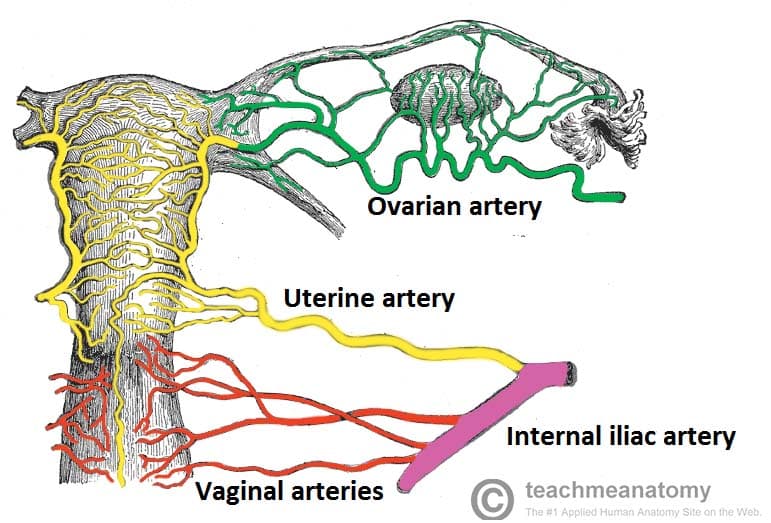
My thought process was that post-partum bleeding is usually related to the uterus, and much of the pelvic viscera is supplied by branches of the internal iliac artery.

get full access to all contentbecome a member
neonem  This sounds like a case of acute endometritis. In any case, uterus is supplied by uterine artery (branch of internal iliac artery) with collateral flow from ovarian artery (comes right off aorta). I don't think there are any branches of external iliac artery into the pelvis; it becomes femoral artery once it passes under inguinal ligament. +4
tsl19  Here's a picture that I found helpful [Female Reproductive Tract arterial supply] (https://teachmeanatomy.info/wp-content/uploads/Blood-Supply-to-Female-Reproductive-Tract.jpg) +18
sympathetikey  @tsl - Thank you! +
step1soon  uworld Qid:11908 +1


submitted by stepwarrior(29), visit this page
get full access to all contentbecome a member

You're just going to diss the son like that right in front of him?

get full access to all contentbecome a member
splanchnic  made an account to upvote this +8
snoodle  I agree - I was stuck between this and "Was he under any unusual stress at home/school or did he have any problems with a gf when all of this started?" and I chose the latter. I didn't want to choose this because why would you want to call the son frightening in front of the son?! I didn't want to choose the answer I chose, either, because you aren't supposed to imply he's straight :/ annoying af +1
makingstrides  I don't think that's what frightening meant. It's meant to describe the mother who is worried about the son. +1


submitted by md_caffeiner(83), visit this page
get full access to all contentbecome a member

what to do with the NONADHERIANT BADDIES???

Intention to treat, "i had the intention to treat so i am gonna leave in this group no matter what"

as treated , "he is not treated as it is so im gonna change his group to control"

per protocol, "you are fired from all of it, protocols bitch"

get full access to all contentbecome a member
johnfred4  I'm just gonna make an Anki cloze card directly from this +
makingstrides  UWORLD QID 19406 Exact question +


submitted by trazobone(97), visit this page
get full access to all contentbecome a member

You would think the rectus femoris is in the back bc rectum, and biceps is in the front. But no. Okay then.

get full access to all contentbecome a member
makingstrides  This is how I remember the biceps, hopefully it helps. Biceps flexes (think of biceps of your arm) - flexion of the leg is raising the leg posteriorly; therefore biceps femoris must be posterior. +2
cweatherly  rectus femoris, I remember ERECTus femoris, because genitalia are in the front +


submitted by pg32(218), visit this page
get full access to all contentbecome a member

Can anyone explain why the lipase concentration is so high if there is an issue with LPL in hyperchylomicronemia?

get full access to all contentbecome a member
garima  due to pancreatitis +7
neovanilla  ELI5? +
suckitnbme  @neovanilla Type 1-hyperchylomicronemia has increased risk of pancreatitis +
makingstrides  I don't understand why this isn't hypertriglyceridemia because this also causes acute pancreatitis. Maybe because overproduction of VLDL isn't an answer? +


submitted by onyx(46), visit this page
get full access to all contentbecome a member

A radiographically visible air-fluid level suggests a pretty large lesion (hence, “cavitary”). That's not going to become normal tissue again. Six months following resolution of symptoms you can expect healing in the form of a scar; that is, fibrosis but only in a single spot.

get full access to all contentbecome a member
masonkingcobra  Robbin's: The basic mechanisms of fibrosis are the same as those of scar formation during tissue repair. However, tissue repair typically occurs after a short-lived injurious stimulus and follows an orderly sequence of steps, whereas fibrosis is induced by persistent injurious stimuli such as infections, immunologic reactions, and other types of tissue injury. The fibrosis seen in chronic diseases such as pulmonary fibrosis is often responsible for organ dysfunction and even organ failure. +5
stevenorange  What Differentiates Normal Lung Repair and Fibrosis? Basement membrane ! https://www.ncbi.nlm.nih.gov/pmc/articles/PMC2645241/ +3
step1234  I'm a little confused here since FA20 209 states Klebsiella causes liquefactive necrosis in the lungs. Does the pulmonary fibrosis occur after the necrosis? +1
fatboyslim  @step1234 Yes pulmonary fibrosis occurs after liquefactive necrosis as a means of repair. Think of it as liquefactive necrosis = damage done in response to Klebsiella -> 6 months later you will see focal fibrosis in that area (scar formation) where the damage happened. +5
makingstrides  If there was an intact basement membrane, you would get normal pulmonary parenchyma. +1
skonys  Not granulomatous inflamm because thats indicative of TB. Not liquefactive nec (trick answer) because Klebsiella causes that actively and q stem says 6Mo post-resolution w/ abx. Not metaplastic pulmonary epithelium because metaplasia implies cellular adaptation to a stimuli (bronchial squamous metaplasia from cigarette smoke), not destruction entirely by liq-nec. IE: for cells to undergo metaplasia they need to be insulted but not destroyed entirely. Not normal pulmonary parenchyma because basement membrane/cytoskeleton architecture is inherently destroyed by cavitary lesion. +3


submitted by hayayah(1212), visit this page
get full access to all contentbecome a member

Neoplasia is new tissue growth that is unregulated, irreversible, and monoclonal.

Clonality can be determined by glucose-6-phosphate dehydrogenase (G6PD) enzyme isoforms. G6PD is X-linked.

*For more information check out Ch. 3 Neoplasia in Pathoma

get full access to all contentbecome a member
hello  This is great, thank you. +5
breis  Pathoma ch. 3 pg 23 "Basic Principles" +9
charcot_bouchard  Shoutout to Imam Satter! Without him this question wasnt possible for me to answer in 10 sec. +19
fatboyslim  Clonality can also be determined by androgen receptor isoforms, which is also present on the X chromosome (Pathoma Ch. 3 Neoplasia) +2
lovebug  @fatboyslim thanks for reminding! +2
makingstrides  Just to make sure I got this right, because this is neoplastic and its monoclonal, you want to look at the isozymes to determine its clonality? +2


submitted by hayayah(1212), visit this page
get full access to all contentbecome a member

Defective homologous recombination is seen in breast/ovarian cancers with the BRCA1 gene mutation.

get full access to all contentbecome a member
johnthurtjr  Ashkenazi Jews have a higher risk of inheriting the BRCA1 and BRCA 2 gene mutations, just another tip! +3
lebron james  BRCA1/BRACA2 are involved in the repair of DNA double stranded breaks +8
samsam3711  Other answers: DNA Mismatch Repair: Lynch Syndrome (MLH1, MSH2) DNA Nucleotide Excision Repair: Xeroderma Pigmentosa +17
lovebug  not about this question but... Defective "Non"-homologous end joining is seen in Ataxia-telangiectasia. :) +4
makingstrides  DNA Base excision repair removed damaged, or not correct, bases. +


submitted by neonem(630), visit this page
get full access to all contentbecome a member

Falling on outstretched hand: scaphoid is most common one to be fractured, lunate is most common to be dislocated. Lunate dislocation can cause acute carpal tunnel syndrome.

Think of the mnemonic "Straight Line To Pinky, Here Comes The Thumb" for the bones of the palm, drawing a football shape starting below the thumb MCP joint adjacent to the radius, then moving to your medial wrist, and then back to the thumb.

Scaphoid, lunate, triquetrum, pisiform, hamate, capitate, trapezoid, trapezium. The lunate looks like it's posteriorly dislocated here.

get full access to all contentbecome a member
sympathetikey  Yep. I didn't even look at the X-ray. +12
dr.xx  loonies love lunate +2
wes79  she landed on her "right hand", but the X-ray is showing a left hand?? +1
wes79  i legit have no idea whats going on in that xray lol +11
nbme4unme  X-ray confused the hell out of me, I was going to put lunate based on Q stem but ended up putting Pisiform because it looks like that's what's messed up in the photo? Should have ignored the picture haha. +1
nwinkelmann  for @dr.xx, love your mnemonic. I added to it, or at least found an explanation on why it works. "loonies love lunate" and "loonies" are "dislocated" from reality. +3
niboonsh  Some Lovers Try Positions That They Cant Handle +13
vsn001  ngl if scaphoid was an option - would've sprung at that real quick -> thanks for teaching me the importance of knowing to look for dislocation vs fracture :D +
regularstudent  Ahh, the classic "left hand" x-ray but actual fracture of "right hand" NBME tactic +
sars  I think the x-ray is showing the lunate protruding out of the palmar side. Imagine the situation where you are falling and using your hand to stop the fall. Your lunate will dislocate forward as the rest of the carpal bones recoil back, hence why it protrudes through the palmar side. Thats why it causes an acute carpal tunnel syndrome. +
makingstrides  Another mnemonic, Some Lovers Tried Positions, That They Can't Handle +
ineedhelpwitmed  so long to pinky, here comes the thumb +
srmtn  in an X Ray (unless is marked) you can not know if is left or right. this is a lunate dislocation but actually is dorsal...even though they put in the stem that is anterior... please google "gilula lines" in order to fastly check a wrist X-ray and find a dislocation... also I think that a good clue when wrist trauma is: dislocation is usually lunate and necrosis is usually scaphoid. hope it helps +


submitted by overa(28), visit this page
get full access to all contentbecome a member

Case-series: comparing patients with a KNOWN OUTCOME (disease) to others with the same disease and looking back to see if they were exposed... "Did 2 people with the same disease get it from the same place?"

Case-control: comparing patients with a KNOWN OUTCOME (disease) to others WITHOUT (control) the disease and looking back to see if they were exposed. (odds ratio)... "they are sick and you want to see if drinking the contaminated water caused it by comparing them to normal people."

Cohort: Comparing patients with a KNOWN EXPOSURE and moving forward with them (prospective) or looking back in time (retrospective) to see if they developed the outcome (disease). (relative risk)... "they all drank the contaminated water and you want to see if they are more likely to get the disease."

Correlation is a statistical analysis used when both the dependent and independent variables have quantitative measurement values.

I hope this is right and helps... :)

get full access to all contentbecome a member
makingstrides  Case series don't have a comparison group . UWorld's answer +


submitted by keycompany(351), visit this page
get full access to all contentbecome a member

This patient has a pneumothorax. Hyperventillation is not enough to compensate for the overall decrease in lung surface area.

get full access to all contentbecome a member
_yeetmasterflex  Could the pneumothorax also cause less ventilation due to decreased lung surface, retaining more CO2 causing respiratory acidosis? That's how I got to the answer at least. +8
duat98  I think pneumothorax would increase RR because you're probably hypoxic. Also I'm sure when you have a lung collapse on you you'd be scared and that would trigger your sympathetic so your RR will go up either way. +4
kateinwonderland  Arterial blood gas studies may show respiratory alkalosis caused by a decrease in CO2 as a result of tachypnea but later hypoxemia, hypercapnia, and acidosis. The patient's SaO2 levels may decrease at first, but typically return to normal within 24 hours. (https://journals.lww.com/nursing/Fulltext/2002/11000/Understanding_pneumothorax.52.aspx) +2
linwanrun1357  How about choice C, --ARDS? +3
bullshitusmle  there is no bilateral lung opacities as you would see in ARDS +5
jesusisking  Was thinking some sort of infection b/c of the atelectasis so picked empyema but this makes sense! +1
djeffs1  does it need to be ARDS to cause "diffuse alveolar damage"? +1
makingstrides  Not only that, does having a collapsed lung affect the alveoli? +1


submitted by lsmarshall(465), visit this page
get full access to all contentbecome a member

Androgen Insensitivity Syndrome - Defect in androgen receptor resulting in normal-appearing female (46,XY DSD). Functioning testes causes increased testosterone at puberty, which is converted to estrogen peripherally, giving female secondary sexual characteristics (female external genitalia). Lack of androgen receptor function leads to absent or scant axillary and pubic hair. Patients have rudimentary vagina, but uterus and fallopian tubes absent.

Androgen insensitivity syndrome is the answer but you might have considered Müllerian agenesis (Mayer-Rokitansky- Küster-Hauser syndrome).

Mullerian agenesis will have normal hormone levels and may present as 1° amenorrhea (due to a lack of uterine development) in females with fully developed 2° sexual characteristics (functional ovaries). Hair development is normal as well. Patients also have normal height.

Seems like this question did not give us much to distinguish besides height and tanner stage 1 pubic/axillary hair.

get full access to all contentbecome a member
dbg  100% agreed. Mullerian agenesis was on my mind too. The full breast development kept me fixed at this dx. Did not think how high testosterone at this age and insensitivity would push towards peripheral conversion to estrogen and hence breast development. Thanks. +
makingstrides  Mullerian agenesis: absent vagina, uterus/cervix because no mullerian system. Yet, still has secondary characteristics ie: breast, pubic hair, normal hormone levels (normal ovaries). Also check to break down the different subtypes of DSD: CAIS, 5alpha reductase deficiency, and swyer syndrome all for XY DSD. Where as for XX DSD, overproduction of gestational androgenism and placental aromatase deficiency. Bc in CAIS the testosterone receptor is dysfunctional, no external / internal male organs are going to form in an XY fetus, but you will have an extra production testosterone (like explained above) leading to increased estrogen (breast growth), but since no ovaries, you dont have the mullerian system. You are left with a vagina with a blind pouch (from lack of functioning receptors) +
makingstrides  Also to add, there are testes that produce the MIH, so you have degeneration of the mullerian system. From B&B +


submitted by guillo12(58), visit this page
get full access to all contentbecome a member

Why there's no increase in Hydrostatic pressure in glomerular capillaries?

get full access to all contentbecome a member
rihan  Between the glomerulus and Bowman's space there is a hydrostatic pressure gradient. This gradient is normally the result of a LARGE glomerular hydrostatic pressure and a LOW pressure in Bowman's space which normally favors filtration. Diagram here: http://physiologyplus.com/wp-content/uploads/2017/08/Glomerular-Filtration.png In the case of post-renal obstruction, hydrostatic pressure behind the blockade will rise and urine will reflux into the capsular space and renal tubules (while glomerular hydrostatic pressure is unaffected) effectively decreasing the pressure gradient which reduces the filtration rate. +6
jurrutia  Why would an increase in pressure in blood vessels cause hydronephrosis? The problem has to be in the nephron itself. +
makingstrides  @jurrutia I think that is being demonstrated here. I picked glomerular capillaries thinking of the answers wrong. The ureteric obstruction causes a backup that leads a to a high hydrostatic pressure in the bowman space. Therefore, preventing filtration. The cause of the renal failure can't be from the glomerular capillaries because the block isn't being caused there; it is caused by something post ureter (dilated ureter). +


search for anything NEW!