Welcome to shervinbd’s page.
Contributor score: 27
Comments ...
Subcomments ...
cassdawg
For me, CMV would have the characteristic "owl eye intranuclear inclusion" cells on biopsy but would be less likely to show anything in pleural fluid (i.e. thoracocentesis would not be used to diagnose CMV). Further CMV pneumonitis is an atypical/interstitial pneumonitis (diffuse patchy infiltrates on CXR, FA2020 p683) and he has a lower lobe consolidation with pleural effusion (more characteristic of fungal pneumonia).
+6
shervinbd
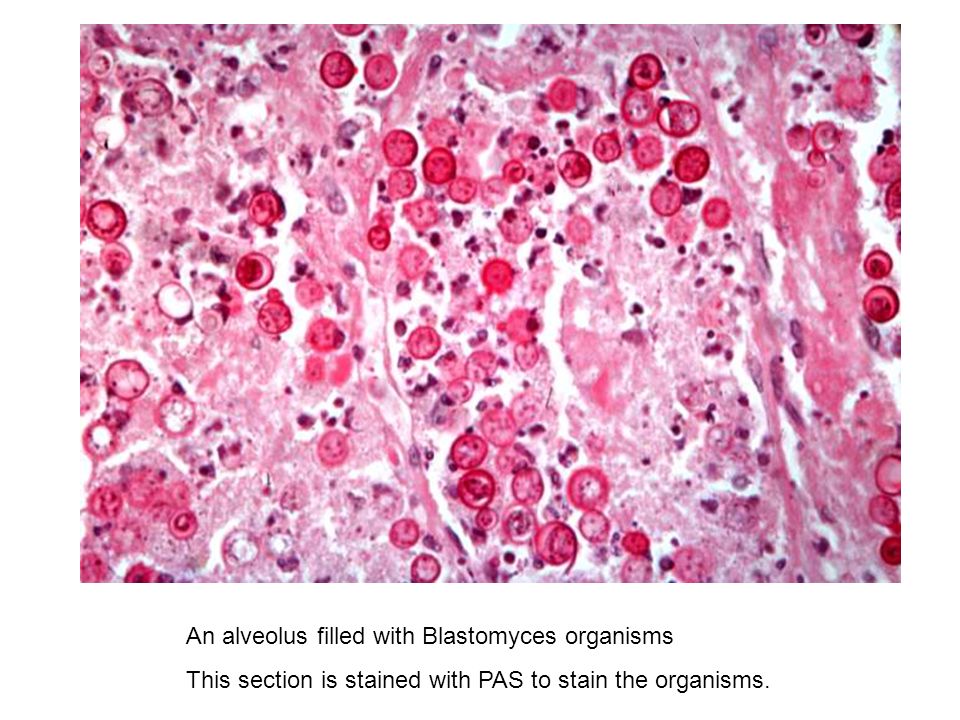
Looks like Cryptococcus neoformans to me.
+4
chaosawaits
I was pretty confident that this is Cryptococcus but the more I look at it, the more I don't know
+1
shervinbd
I think it is Ascaris, not Stringlyloides. The symptoms could be explained by Loeffler syndrome, caused by Ascaris larva migration. Ascaris is transmitted through fecal oral route, so ingestion of feces contaminated soil could cause the problem. Per FA, Strongylides is transmitted by larva penetrating skin.
+23
drmifta
Its Ascaris. Fecal oral transmission -> Larvae penitrate GIT -> Blood Stream -> Lung {Maturation, Respiratory Symptom} -> Coughed up and swallowing -> Adult Warm in GIT -> Egg release -> Egg in stool.
+1
i_hate_it_here
Didn't the stem mention that roundwarm larvae were found? I thought Ascaris is diagnosed by bile coated eggs in feces?
+1
sexymexican888
I actually think @cassdawg is right. Its strongyloides. They found larvae in the feces (you find eggs in feces with ascaris) you can get pulmonary sx in both. Ascaris is also usually fecal oral transmission so its more likely to come from someone making food with contaminated hands. Strongyloides is transmitted through soil or sand and the larvae penetrate your feet so this makes more sense.
+2
sexymexican888
You can find this is FA 2020 Pg 159. Also if you look at the table strongyloides is assoc. with pulmonary sx. However I think its both cause according to sketchy micro ascaris presents with respiratory sx
+
coco
Although Strongyloides nematode worm infections are not overly common in the United States, the Appalachian area of the Southeast have reported cases.
Ascaris lumbricoides is a common parasitic infection in Asia, Africa, and South America. Most cases in the United States arise in travelers to these regions.
Loeffler syndrome can be caused by Strongyloides and Ascaris.
Ascar:Stool microscopy reveals an oval egg with a thick outer shell and a single interior ovum
Strongyloides:rhabditiform larvae seen in feces under microscope
so.I think this is Strongyloides.If I make a mistake,please correct me.
+2
melanoma
I think is ascaris lumbricoides also due to the size of the larva. UW Q 15549
+
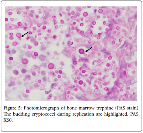
Can someone please explain what we're seeing on the histo slide? I chose the correct answer because I was thinking fungus because of the immunocompromise and neutropenia (and I thought PAS was used for aspergillus), but I don't see anything fungus-related on that slide.